 Explore
 Validate
 Learn
Learn Western blot
Antibody data
- Antibody Data
- Antigen structure
- References [2]
- Comments [0]
- Validations
- Western blot [5]
- Immunocytochemistry [1]
- Immunoprecipitation [6]
- Chromatin Immunoprecipitation [3]
Submit
Validation data
Reference
Comment
Report error
- Product number
- GTX121945 - Provider product page

- Provider
- GeneTex
- Proper citation
- GeneTex Cat#GTX121945, RRID:AB_10721057
- Product name
- SMYD3 antibody
- Antibody type
- Polyclonal
- Reactivity
- Human, Mouse
- Host
- Rabbit
Submitted references SMYD3 Promotes Homologous Recombination via Regulation of H3K4-mediated Gene Expression.
The methyltransferase SMYD3 mediates the recruitment of transcriptional cofactors at the myostatin and c-Met genes and regulates skeletal muscle atrophy.
Chen YJ, Tsai CH, Wang PY, Teng SC
Scientific reports 2017 Jun 19;7(1):3842
Scientific reports 2017 Jun 19;7(1):3842
The methyltransferase SMYD3 mediates the recruitment of transcriptional cofactors at the myostatin and c-Met genes and regulates skeletal muscle atrophy.
Proserpio V, Fittipaldi R, Ryall JG, Sartorelli V, Caretti G
Genes & development 2013 Jun 1;27(11):1299-312
Genes & development 2013 Jun 1;27(11):1299-312
No comments: Submit comment
Supportive validation
- Submitted by
- GeneTex (provider)
- Main image
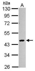
- Experimental details
- Sample (30 ug of whole cell lysate) A: 293T 10% SDS PAGE GTX121945 diluted at 1:1000
- Validation comment
- WB
- Submitted by
- GeneTex (provider)
- Main image
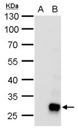
- Experimental details
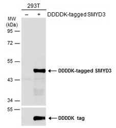
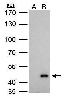
- SMYD3 antibody detects SMYD3 protein by western blot analysis.A. 30 £gg 293T whole cell lysate/extract B. 30 £gg whole cell lysate/extract of human SMYD3-transfected 293T cells (partial fragment)10 % SDS-PAGESMYD3 antibody (GTX121945) dilution: 1:1000

- Submitted by
- GeneTex (provider)
- Main image
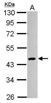
- Experimental details
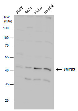
- SMYD3 antibody detects SMYD3 protein by western blot analysis. Various whole cell extracts (30 £gg) were separated by 10% SDS-PAGE, and the membrane was blotted with SMYD3 antibody (GTX121945) diluted at a dilution of 1:1000.
- Submitted by
- GeneTex (provider)
- Main image
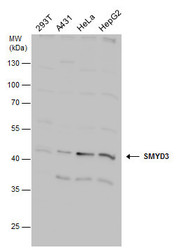
- Experimental details
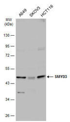
- Various whole cell extracts (30 ?g) were separated by 10% SDS-PAGE, and the membrane was blotted with SMYD3 antibody (GTX121945) diluted at 1:1000. The HRP-conjugated anti-rabbit IgG antibody (GTX213110-01) was used to detect the primary antibody.
- Submitted by
- GeneTex (provider)
- Main image
- Experimental details
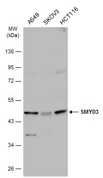
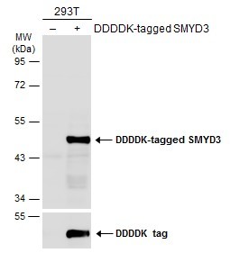
- Non-transfected (¡V) and transfected (+) 293T whole cell extracts (30 ?g) were separated by 10% SDS-PAGE, and the membrane was blotted with SMYD3 antibody (GTX121945) diluted at 1:1000. The HRP-conjugated anti-rabbit IgG antibody (GTX213110-01) was used to detect the primary antibody.
Supportive validation
- Submitted by
- GeneTex (provider)
- Main image
- Experimental details
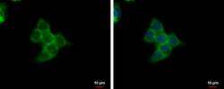
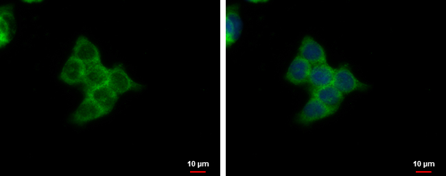
- SMYD3 antibody detects SMYD3 protein at cytoplasm by immunofluorescent analysis.Sample: HCT116 cells were fixed in ice-cold MeOH for 5 min.Green: SMYD3 protein stained by SMYD3 antibody (GTX121945) diluted at 1:500.Blue: Hoechst 33342 staining.
Supportive validation
- Submitted by
- GeneTex (provider)
- Main image
- Experimental details
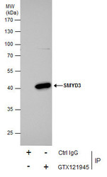
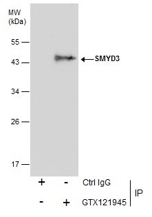
- SMYD3 antibody immunoprecipitates SMYD3 protein in IP experiments. IP Sample: 293T whole cell lysate/extract A. Control with 2 ?g of preimmune rabbit IgG B. Immunoprecipitation of SMYD3 protein by 2 ?g of SMYD3 antibody (GTX121945) 7.5% SDS-PAGE The immunoprecipitated SMYD3 protein was detected by SMYD3 antibody (GTX121945) diluted at 1:1000. EasyBlot anti-rabbit IgG (GTX221666-01) was used as a secondary reagent.
- Validation comment
- IP
- Submitted by
- GeneTex (provider)
- Main image
- Experimental details
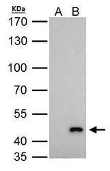
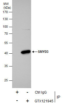
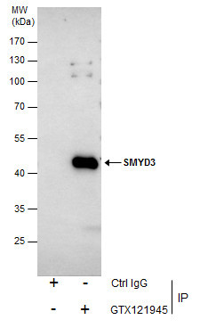
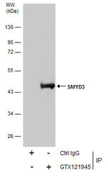
- Immunoprecipitation of SMYD3 protein from 293T whole cell extracts using 5 ?g of SMYD3 antibody (GTX121945).Western blot analysis was performed using SMYD3 antibody (GTX121945).EasyBlot anti-Rabbit IgG (GTX221666-01) was used as a secondary reagent.
- Validation comment
- IP
- Submitted by
- GeneTex (provider)
- Main image
- Experimental details
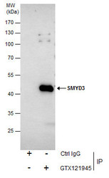
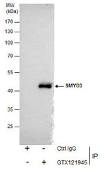
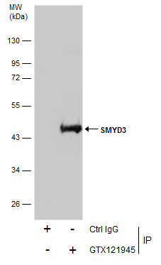
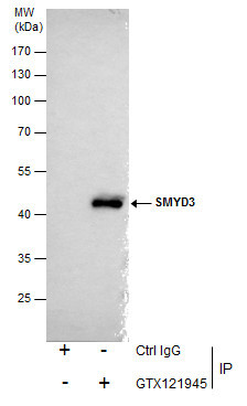
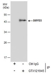
- Immunoprecipitation of SMYD3 protein from 293T whole cell extracts using 5 £gg of SMYD3 antibody (GTX121945).Western blot analysis was performed using SMYD3 antibody (GTX121945).EasyBlot anti-Rabbit IgG (GTX221666-01) was used as a secondary reagent.
- Submitted by
- GeneTex (provider)
- Main image
- Experimental details
- Immunoprecipitation of SMYD3 protein from 293T whole cell extracts using 5 £gg of SMYD3 antibody (GTX121945).Western blot analysis was performed using SMYD3 antibody (GTX121945).EasyBlot anti-Rabbit IgG (GTX221666-01) was used as a secondary reagent.
Supportive validation
- Submitted by
- GeneTex (provider)
- Main image

- Experimental details
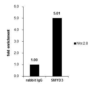
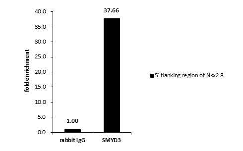
- Cross-linked ChIP was performed with HepG2 chromatin extract and 5 ?g of either control rabbit IgG or anti-SMYD3 antibody. The precipitated DNA was detected by PCR with primer set targeting to 5' flanking region of Nkx2.8.
- Validation comment
- ChIP
- Submitted by
- GeneTex (provider)
- Main image

- Experimental details
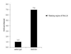
- Cross-linked ChIP was performed with HepG2 chromatin extract and 5 ?g of either normal rabbit IgG or anti-SMYD3 antibody. The precipitated DNA was detected by PCR with primer set targeting to 5' flanking region of Nkx2.8.
- Validation comment
- ChIP
- Submitted by
- GeneTex (provider)
- Main image
- Experimental details
- Cross-linked ChIP was performed with HepG2 chromatin extract and 5 £gg of either normal rabbit IgG or anti-SMYD3 antibody. The precipitated DNA was detected by PCR with primer set targeting to 5' flanking region of Nkx2.8..
