Explore
 Validate
 Learn
Learn Western blot
Antibody data
- Antibody Data
- Antigen structure
- References [0]
- Comments [0]
- Validations
- Western blot [1]
- Immunocytochemistry [1]
- Immunohistochemistry [1]
- Flow cytometry [1]
Submit
Validation data
Reference
Comment
Report error
- Product number
- GTX16925 - Provider product page

- Provider
- GeneTex
- Product name
- P2X1 antibody
- Antibody type
- Polyclonal
- Reactivity
- Human, Mouse, Rat
- Host
- Rabbit
No comments: Submit comment
Supportive validation
- Submitted by
- GeneTex (provider)
- Main image

- Experimental details
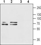
- Anti-P2X1_Receptor_(extracellular) - Western blot analysis of rat brain (lanes 1 and 3) and mouse brain (lanes 2 and 4) lysates: 1, 2. Anti-P2X1 Receptor (extracellular) antibody, (1:200). 3, 4. Anti-P2X1 Receptor (extracellular) antibody, preincubated with the control peptide antigen.
Supportive validation
- Submitted by
- GeneTex (provider)
- Main image
- Experimental details
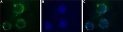
- Anti-P2X1_Receptor_(extracellular) - Expression of P2X1 in human SH-SYS5 cells Immunocytochemical staining of intact living human SH-SYS5 cells. A. Extracellular staining of cells with Anti-P2X1 Receptor (extracellular) antibody, (1:50) followed by goat anti-rabbit-AlexaFluor-488 secondary antibody. B. Nuclear staining DAPI as the counterstain. C. Merged images of A and B.
Supportive validation
- Submitted by
- GeneTex (provider)
- Main image
- Experimental details
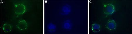
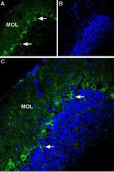
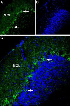
- Anti-P2X1_Receptor_(extracellular) - Expression of P2X1 in mouse cerebellum Immunohistochemical staining of mouse cerebellum using Anti-P2X1 Receptor (extracellular) antibody. A. Most of P2X1 labeling (green) appears in fine processes in the molecular layer (MOL) and in Purkinje cells (arrows show examples). B. DAPI is used as the counterstain (blue). C. Merge of A and B.
Supportive validation
- Submitted by
- GeneTex (provider)
- Main image
- Experimental details
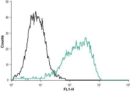
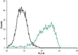
- Anti-P2X1_Receptor_(extracellular) - Indirect flow cytometry analysis of MEG-O1 cells: ___ Unstained cells.___ Cells + Anti-P2X1 Receptor (extracellular) antibody, (10 £gg antibody/1x106 cells).