 Explore
 Validate
 Learn
Learn Western blot
Antibody data
- Antibody Data
- Antigen structure
- References [0]
- Comments [0]
- Validations
- Western blot [1]
- Immunohistochemistry [2]
Submit
Validation data
Reference
Comment
Report error
- Product number
- TA329025 - Provider product page

- Provider
- OriGene
- Product name
- Rabbit polyclonal Anti-Ryanodine Receptor 2
- Antibody type
- Polyclonal
- Description
- Rabbit polyclonal Anti-Ryanodine Receptor 2
- Host
- Rabbit
- Conjugate
- Unconjugated
- Epitope
- RYR2
- Antibody clone number
- NULL
- Vial size
- 200 µl
- Concentration
- NULL
No comments: Submit comment
Supportive validation
- Submitted by
- OriGene (provider)
- Main image
- Experimental details
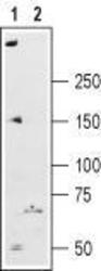
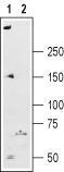
- Western blot analysis of rat heart membranes: 1. Anti-Ryanodine Receptor 2 antibody , (1:200). 2. Anti-Ryanodine Receptor 2 antibody, preincubated with the control peptide antigen.
- Validation comment
- WB
Supportive validation
- Submitted by
- OriGene (provider)
- Main image

- Experimental details
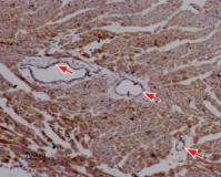
- Expression of Ryanodine Receptor 2 in rat cardiac muscle. Immunohistochemical staining of paraffin-embedded sections of rat myocardium using Anti-Ryanodine Receptor 2 antibody , (1:50). Staining is specific for cardiomyocytes while smooth muscles cells in the artery walls are negative (red arrows). Hematoxilin is used as the counterstain.
- Validation comment
- IHC
- Submitted by
- OriGene (provider)
- Main image
- Experimental details
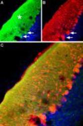
- IHC staining of mouse cerebellum frozen sections with Anti-Ryanodine Receptor 2 antibody , (1:100), (green). A. The highest expression of Ryanodine Receptor 2 is in the molecular layer (Asterisk) but there is also some expression in the soma of Purkinje cells (arrows). B. In the same section, there is staining for parvalbumin (red). C. Merged image of panels A and B demonstrates Ryanodine Receptor 2 is localized both in the area surrounding the dendritic tree and in the soma of Purkinje cells.
- Validation comment
- IHC
