 Explore
 Validate
 Learn
Learn12-5129-42
antibody from Invitrogen Antibodies
Targeting: LILRB1
CD85, CD85j, ILT2, LIR-1, LIR1, MIR-7, PIR-B, PIRB
 Flow cytometry
Antibody data
- Antibody Data
- Antigen structure
- References [7]
- Comments [0]
- Validations
- Flow cytometry [2]
- Other assay [3]
Submit
Validation data
Reference
Comment
Report error
- Product number
- 12-5129-42 - Provider product page

- Provider
- Invitrogen Antibodies
- Product name
- CD85j (ILT2) Monoclonal Antibody (HP-F1), PE, eBioscience™
- Antibody type
- Monoclonal
- Antigen
- Other
- Description
- Description: The monoclonal antibody HP-F1 recognizes CD85j, also known as ILT2, LILRB1, and LIR1. CD85j is a member of the ILT (immunoglobulin-like transcript)/LIR (leukocyte Ig-like receptor)/MIR (monocyte Ig-like receptor) family. CD85j is a single transmembrane glycoprotein with a long cytoplasmic domain containing 4 ITIMs which signal through interactions with SHP-1. Expression is found on myeloid cells (monocytes and dendritic cells) and some lymphoid cells including, subsets of NK, T and B cells. Expression has been correlated with leukemias such as ALL and CLL. Expression on CD8+ cells correlates with effector cell function and plays an important role in viral infections, including HIV, Ebstein Barr and CMV. The ligands for CD85j are MHC Class I molecules such as HLA-G, A, F, B27, E and F. The monoclonal antibody HP-F1 has been shown to reduce the amount of CD16- dependent cytolytic activity of functional NK cells. Applications Reported: This HP-F1 antibody has been reported for use in flow cytometric analysis. Applications Tested: This HP-F1 antibody has been pre-titrated and tested by flow cytometric analysis of normal human peripheral blood cells. This can be used at 5 µL (0.125 µg) per test. A test is defined as the amount (µg) of antibody that will stain a cell sample in a final volume of 100 µL. Cell number should be determined empirically but can range from 10^5 to 10^8 cells/test. Excitation: 488-561 nm; Emission: 578 nm; Laser: Blue Laser, Green Laser, Yellow-Green Laser. Filtration: 0.2 µm post-manufacturing filtered.
- Reactivity
- Human
- Host
- Mouse
- Conjugate
- Yellow dye
- Isotype
- IgG
- Antibody clone number
- HP-F1
- Vial size
- 100 Tests
- Concentration
- 5 μL/Test
- Storage
- 4°C, store in dark, DO NOT FREEZE!
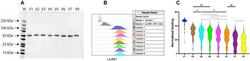
Submitted references Natural LILRB1 D1-D2 variants show frequency differences in populations and bind to HLA class I with various avidities.
Interaction between HLA-G and NK cell receptor KIR2DL4 orchestrates HER2-positive breast cancer resistance to trastuzumab.
Ig-Like Transcript 2 (ILT2) Blockade and Lenalidomide Restore NK Cell Function in Chronic Lymphocytic Leukemia.
Prediction of non-muscle-invasive bladder cancer recurrence by measurement of checkpoint HLAG's receptor ILT2 on peripheral CD8(+) T cells.
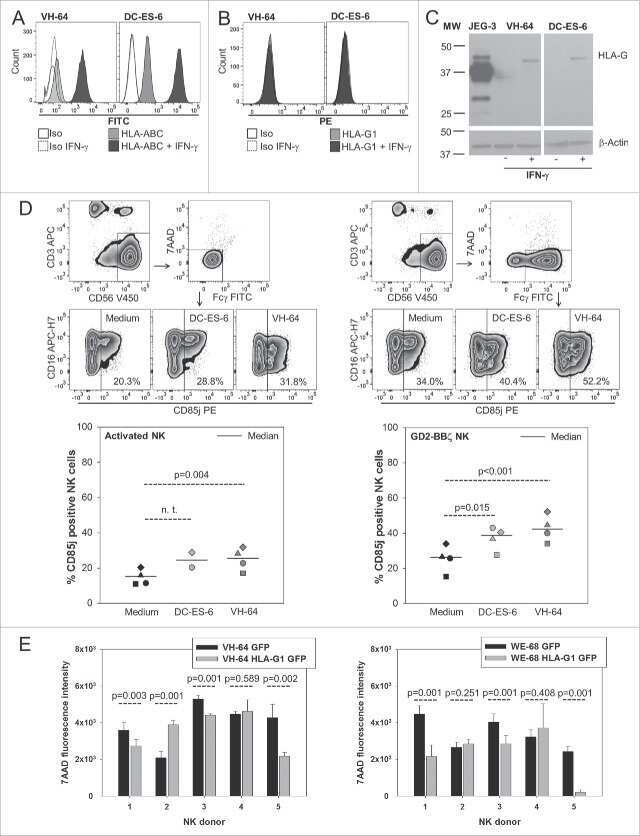
Targeting Ewing sarcoma with activated and GD2-specific chimeric antigen receptor-engineered human NK cells induces upregulation of immune-inhibitory HLA-G.
Identification of GAD65 AA 114-122 reactive 'memory-like' NK cells in newly diagnosed Type 1 diabetic patients by HLA-class I pentamers.
A common inhibitory receptor for major histocompatibility complex class I molecules on human lymphoid and myelomonocytic cells.
Liu F, Cocker ATH, Pugh JL, Djaoud Z, Parham P, Guethlein LA
Immunogenetics 2022 Dec;74(6):513-525
Immunogenetics 2022 Dec;74(6):513-525
Interaction between HLA-G and NK cell receptor KIR2DL4 orchestrates HER2-positive breast cancer resistance to trastuzumab.
Zheng G, Guo Z, Li W, Xi W, Zuo B, Zhang R, Wen W, Yang AG, Jia L
Signal transduction and targeted therapy 2021 Jun 23;6(1):236
Signal transduction and targeted therapy 2021 Jun 23;6(1):236
Ig-Like Transcript 2 (ILT2) Blockade and Lenalidomide Restore NK Cell Function in Chronic Lymphocytic Leukemia.
Villa-Álvarez M, Sordo-Bahamonde C, Lorenzo-Herrero S, Gonzalez-Rodriguez AP, Payer AR, Gonzalez-Garcia E, Villa-Álvarez MC, López-Soto A, Gonzalez S
Frontiers in immunology 2018;9:2917
Frontiers in immunology 2018;9:2917
Prediction of non-muscle-invasive bladder cancer recurrence by measurement of checkpoint HLAG's receptor ILT2 on peripheral CD8(+) T cells.
Desgrandchamps F, LeMaoult J, Goujon A, Riviere A, Rivero-Juarez A, Djouadou M, de Gouvello A, Dumont C, Wu CL, Culine S, Verine J, Rouas-Freiss N, Hennequin C, Masson-Lecomte A, Carosella ED
Oncotarget 2018 Sep 4;9(69):33160-33169
Oncotarget 2018 Sep 4;9(69):33160-33169
Targeting Ewing sarcoma with activated and GD2-specific chimeric antigen receptor-engineered human NK cells induces upregulation of immune-inhibitory HLA-G.
Kailayangiri S, Altvater B, Spurny C, Jamitzky S, Schelhaas S, Jacobs AH, Wiek C, Roellecke K, Hanenberg H, Hartmann W, Wiendl H, Pankratz S, Meltzer J, Farwick N, Greune L, Fluegge M, Rossig C
Oncoimmunology 2017;6(1):e1250050
Oncoimmunology 2017;6(1):e1250050
Identification of GAD65 AA 114-122 reactive 'memory-like' NK cells in newly diagnosed Type 1 diabetic patients by HLA-class I pentamers.
Perri V, Gianchecchi E, Cifaldi L, Pellegrino M, Giorda E, Andreani M, Cappa M, Fierabracci A
PloS one 2017;12(12):e0189615
PloS one 2017;12(12):e0189615
A common inhibitory receptor for major histocompatibility complex class I molecules on human lymphoid and myelomonocytic cells.
Colonna M, Navarro F, Bellón T, Llano M, García P, Samaridis J, Angman L, Cella M, López-Botet M
The Journal of experimental medicine 1997 Dec 1;186(11):1809-18
The Journal of experimental medicine 1997 Dec 1;186(11):1809-18
No comments: Submit comment
Supportive validation
- Submitted by
- Invitrogen Antibodies (provider)
- Main image
- Experimental details
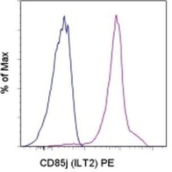
- Staining of normal human peripheral blood cells with Mouse IgG1 K Isotype Control PE (Product # 12-4714-81) (blue histogram) or Anti-Human CD85j (ILT2) PE (purple histogram). Cells in the monocyte gate were used for analysis.
- Conjugate
- Yellow dye
- Submitted by
- Invitrogen Antibodies (provider)
- Main image
- Experimental details
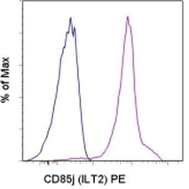
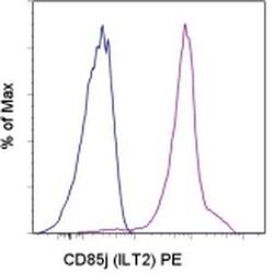
- Staining of normal human peripheral blood cells with Mouse IgG1 K Isotype Control PE (Product # 12-4714-81) (blue histogram) or Anti-Human CD85j (ILT2) PE (purple histogram). Cells in the monocyte gate were used for analysis.

Supportive validation
- Submitted by
- Invitrogen Antibodies (provider)
- Main image
- Experimental details
- NULL
- Conjugate
- Yellow dye
- Submitted by
- Invitrogen Antibodies (provider)
- Main image

- Experimental details
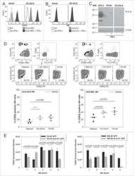
- 10.1371/journal.pone.0189615.g004 Fig 4 Similar percentage of total and ILT2 expressing NK cells between healthy donors and T1D patients. The percentage of CD3 - CD56 + cells (A) and of CD3 - CD56 + ILT2 + cells (B) were assessed by flow cytometric analyses of IL-2 alone treated PBMC. NK cell phenotype of 14 healthy donors (HD, circle plots) was compared with that of 14 T1D patients (square dots); horizontal bars, average values are shown. No significant differences are reported (KS test, unpaired t test).
- Conjugate
- Yellow dye

- Submitted by
- Invitrogen Antibodies (provider)
- Main image
- Experimental details
- Fig. 4 LILRB1 D1-D2 variants bind to HLA class I with different avidities. A SDS-PAGE analysis of the eight successfully produced and purified LILRB1-D1D2-Fc proteins. B Representative integrity analysis of the D1-D2 portion of the produced proteins by flow cytometry using anti-human LILRB1 antibody. C Violin plots of the binding data of each of LILRB1 D1-D2 variants to 97 combined HLA class I tested by single antigen bead assay. The dashed line indicates there is no significant difference between variants, the single star indicates p
- Conjugate
- Yellow dye
