Explore
 Validate
 Learn
Learn Western blot
Antibody data
- Antibody Data
- Antigen structure
- References [1]
- Comments [0]
- Validations
- Western blot [6]
- Immunocytochemistry [2]
- Immunohistochemistry [3]
Submit
Validation data
Reference
Comment
Report error
- Product number
- GTX114394 - Provider product page

- Provider
- GeneTex
- Proper citation
- GeneTex Cat#GTX114394, RRID:AB_10619907
- Product name
- ERM antibody
- Antibody type
- Polyclonal
- Reactivity
- Human, Mouse
- Host
- Rabbit
Submitted references Maldevelopment of the submandibular gland in a mouse model of apert syndrome.
Yamaji K, Morita J, Watanabe T, Gunjigake K, Nakatomi M, Shiga M, Ono K, Moriyama K, Kawamoto T
Developmental dynamics : an official publication of the American Association of Anatomists 2018 Nov;247(11):1175-1185
Developmental dynamics : an official publication of the American Association of Anatomists 2018 Nov;247(11):1175-1185
No comments: Submit comment
Supportive validation
- Submitted by
- GeneTex (provider)
- Main image
- Experimental details
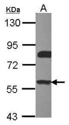
- Sample (30 ug of whole cell lysate) A: Raji 7.5% SDS PAGE ERM antibody GTX114394 diluted at 1:1000
- Validation comment
- WB

- Submitted by
- GeneTex (provider)
- Main image

- Experimental details
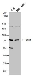
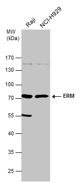
- ERM antibody detects ERM protein by western blot analysis.A. 30 ?g Raji whole cell lysate/extractB. 30 ?g NCI-H929 whole cell lysate/extract7.5 % SDS-PAGEERM antibody (GTX114394) dilution: 1:500
- Validation comment
- WB
- Submitted by
- GeneTex (provider)
- Main image
- Experimental details
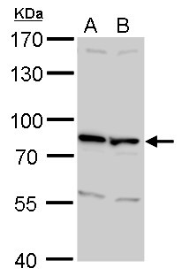
- Various whole cell extracts (30 ?g) were separated by 10% SDS-PAGE, and the membrane was blotted with ERM antibody (GTX114394) diluted at 1:500. The HRP-conjugated anti-rabbit IgG antibody (GTX213110-01) was used to detect the primary antibody.
- Submitted by
- GeneTex (provider)
- Main image

- Experimental details
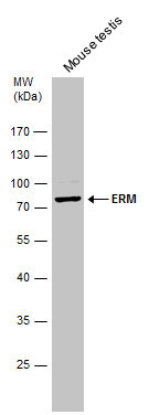
- Mouse tissue extract (50 ?g) was separated by 10% SDS-PAGE, and the membrane was blotted with ERM antibody (GTX114394) diluted at 1:500. The HRP-conjugated anti-rabbit IgG antibody (GTX213110-01) was used to detect the primary antibody.
- Submitted by
- GeneTex (provider)
- Main image
- Experimental details
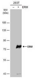
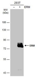
- Non-transfected (¡V) and transfected (+) 293T whole cell extracts (30 ?g) were separated by 10% SDS-PAGE, and the membrane was blotted with ERM antibody (GTX114394) diluted at 1:5000. The HRP-conjugated anti-rabbit IgG antibody (GTX213110-01) was used to detect the primary antibody.
- Submitted by
- GeneTex (provider)
- Main image
- Experimental details
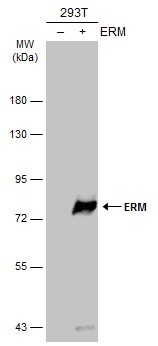
- Non-transfected (¡V) and transfected (+) 293T whole cell extracts (30 ?g) were separated by 10% SDS-PAGE, and the membrane was blotted with ERM antibody (GTX114394) diluted at 1:5000. The HRP-conjugated anti-rabbit IgG antibody (GTX213110-01) was used to detect the primary antibody.

Supportive validation
- Submitted by
- GeneTex (provider)
- Main image

- Experimental details
- Immunofluorescence analysis of paraformaldehyde-fixed A431, using ERM(GTX114394) antibody at 1:500 dilution.

- Submitted by
- GeneTex (provider)
- Main image
- Experimental details
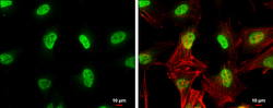
- ERM antibody detects ERM protein at nucleus by immunofluorescent analysis.Sample: HeLa cells were fixed in 4% paraformaldehyde at RT for 15 min.Green: ERM protein stained by ERM antibody (GTX114394) diluted at 1:500.Red: Phalloidin, a cytoskeleton marker, diluted at 1:100.Blue: Hoechst 33342 staining.Scale bar = 10 £gm.

Supportive validation
- Submitted by
- GeneTex (provider)
- Main image

- Experimental details
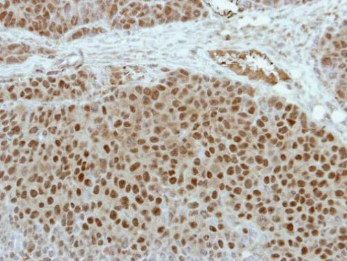
- Immunohistochemical analysis of paraffin-embedded PC14 xenograft, using ERM(GTX114394) antibody at 1:100 dilution.
- Submitted by
- GeneTex (provider)
- Main image

- Experimental details
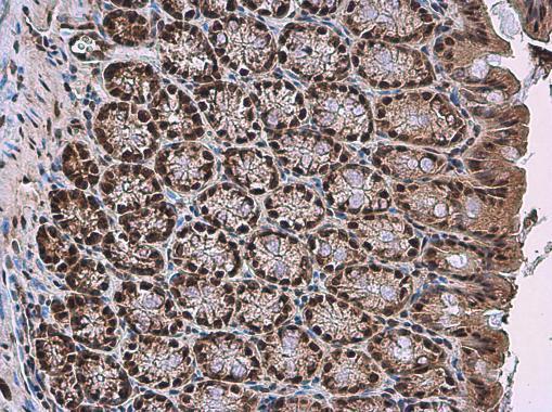
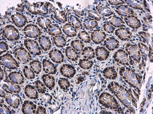
- ERM antibody detects ERM protein at nucleus in mouse colon by immunohistochemical analysis. Sample: Paraffin-embedded mouse colon. ERM antibody (GTX114394) diluted at 1:500.
- Submitted by
- GeneTex (provider)
- Main image
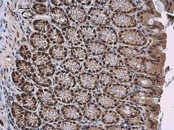
- Experimental details
- ERM antibody detects ERM protein at nucleus in mouse colon by immunohistochemical analysis. Sample: Paraffin-embedded mouse colon. ERM antibody (GTX114394) diluted at 1:500.