 Explore
 Validate
 Learn
Learn Western blot
Antibody data
- Antibody Data
- Antigen structure
- References [2]
- Comments [0]
- Validations
- Western blot [3]
Submit
Validation data
Reference
Comment
Report error
- Product number
- AB-289-PB - Provider product page

- Provider
- Novus Biologicals
- Product name
- Goat Polyclonal MIF Antibody
- Antibody type
- Polyclonal
- Description
- Protein A or G purified. Detects human MIF in Western blots. Because this antibody preparation is a total IgG fration, complete monospecificity cannot be assumed.
- Reactivity
- Human, Mouse
- Host
- Goat
- Conjugate
- Unconjugated
- Isotype
- IgG
- Vial size
- 1 mg
- Concentration
- LYOPH
- Storage
- Use a manual defrost freezer and avoid repeated freeze-thaw cycles. 12 months from date of receipt, -20 to -70 degreesC as supplied. 1 month, 2 to 8 degreesC under sterile conditions after reconstitution. 6 months, -20 to -70 degreesC under sterile conditions after reconstitution.
Submitted references The sex steroid precursor DHEA accelerates cutaneous wound healing via the estrogen receptors.
Estrogen modulates cutaneous wound healing by downregulating macrophage migration inhibitory factor.
Mills SJ, Ashworth JJ, Gilliver SC, Hardman MJ, Ashcroft GS
The Journal of investigative dermatology 2005 Nov;125(5):1053-62
The Journal of investigative dermatology 2005 Nov;125(5):1053-62
Estrogen modulates cutaneous wound healing by downregulating macrophage migration inhibitory factor.
Ashcroft GS, Mills SJ, Lei K, Gibbons L, Jeong MJ, Taniguchi M, Burow M, Horan MA, Wahl SM, Nakayama T
The Journal of clinical investigation 2003 May;111(9):1309-18
The Journal of clinical investigation 2003 May;111(9):1309-18
No comments: Submit comment
Supportive validation
- Submitted by
- Novus Biologicals (provider)
- Main image

- Experimental details
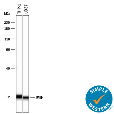
- Detection of Human MIF by Simple WesternTM. Simple Western lane view shows lysates of THP-1 human acute monocytic leukemia cell line and U937 human histiocytic lymphoma cell line, loaded at 0.2 mg/mL. A specific band was detected for MIF at approximately 12 kDa (as indicated) using 25 µg/mL of Goat Anti-Human/Mouse MIF Polyclonal Antibody (Catalog # AB-289-PB) followed by 1:50 dilution of HRP-conjugated Anti-Goat IgG Secondary Antibody (Catalog # HAF109). This experiment was conducted under reducing conditions and using the 12-230 kDa separation system.
- Submitted by
- Novus Biologicals (provider)
- Main image

- Experimental details
- Detection of Human MIF by Western Blot. Western blot shows lysates of THP-1 human acute monocytic leukemia cell line and U937 human histiocytic lymphoma cell line. PVDF membrane was probed with 0.2 µg/mL of Goat Anti-Human/Mouse MIF Polyclonal Antibody (Catalog # AB-289-PB) followed by HRP-conjugated Anti-Goat IgG Secondary Antibody (Catalog # HAF017). A specific band was detected for MIF at approximately 12 kDa (as indicated). This experiment was conducted under reducing conditions and using Immunoblot Buffer Group 1.

- Submitted by
- Novus Biologicals (provider)
- Main image
- Experimental details
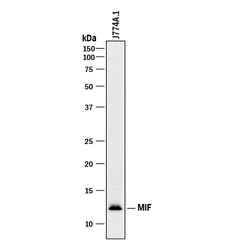
- Detection of Mouse MIF by Western Blot. Western blot shows lysates of J774A.1 mouse reticulum cell sarcoma macrophage cell line. PVDF membrane was probed with 1 µg/mL of Goat Anti-Human/Mouse MIF Polyclonal Antibody (Catalog # AB-289-PB) followed by HRP-conjugated Anti-Goat IgG Secondary Antibody (Catalog # HAF017). A specific band was detected for MIF at approximately 12 kDa (as indicated). This experiment was conducted under reducing conditions and using Immunoblot Buffer Group 1.
