 Explore
 Validate
 Learn
Learn Western blot
Antibody data
- Antibody Data
- Antigen structure
- References [0]
- Comments [0]
- Validations
- Western blot [1]
- Flow cytometry [1]
Submit
Validation data
Reference
Comment
Report error
- Product number
- MAB2891 - Provider product page

- Provider
- Novus Biologicals
- Product name
- Mouse Monoclonal MIF Antibody
- Antibody type
- Monoclonal
- Description
- Protein A or G purified from hybridoma culture supernatant. Detects human MIF in direct ELISAs.
- Reactivity
- Human
- Host
- Mouse
- Conjugate
- Unconjugated
- Isotype
- IgG
- Vial size
- 100 ug
- Storage
- Use a manual defrost freezer and avoid repeated freeze-thaw cycles. 12 months from date of receipt, -20 to -70 degreesC as supplied. 1 month, 2 to 8 degreesC under sterile conditions after reconstitution. 6 months, -20 to -70 degreesC under sterile conditions after reconstitution.
No comments: Submit comment
Supportive validation
- Submitted by
- Novus Biologicals (provider)
- Main image

- Experimental details
- Detection of Human MIF by Western Blot. Western blot shows lysates of THP-1 human acute monocytic leukemia cell line and U937 human histiocytic lymphoma cell line. PVDF membrane was probed with 2 µg/mL of Mouse Anti-Human MIF Monoclonal Antibody (Catalog # MAB2891) followed by HRP-conjugated Anti-Mouse IgG Secondary Antibody (Catalog # HAF018). A specific band was detected for MIF at approximately 12 kDa (as indicated). This experiment was conducted under reducing conditions and using Immunoblot Buffer Group 1. It is recommended to use Mouse Anti-Human MIF Monoclonal Antibody (Catalog # MAB2893, Clone # 932603) as an alternative antibody for Western blot.

Supportive validation
- Submitted by
- Novus Biologicals (provider)
- Main image
- Experimental details
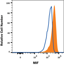
- Detection of MIF in Human PBMCs by Flow Cytometry. Human peripheral blood mononuclear cell (PBMCs), resting (open histogram), or treated with 1 ug/mL LPS overnight and 3 uM monensin for 2 hours (filed histogram) were stained with Mouse Anti-Human MIF Monoclonal Antibody (Catalog # MAB2891) or isotype control antibody (Catalog # MAB002), followed by Phycoerythrin-conjugated Anti-Mouse IgG Secondary Antibody (Catalog # F0102B). To facilitate intracellular staining, cells were fixed with Flow Cytometry Fixation Buffer (Catalog # FC004) and permeabilized with Flow Cytometry Permeabilization/Wash Buffer I (Catalog # FC005).
