Explore
 Validate
 Learn
Learn Western blot
Antibody data
- Antibody Data
- Antigen structure
- References [1]
- Comments [0]
- Validations
- Western blot [5]
- Immunoprecipitation [1]
- Immunohistochemistry [3]
Submit
Validation data
Reference
Comment
Report error
- Product number
- NBP2-20398 - Provider product page

- Provider
- Novus Biologicals
- Product name
- Rabbit Polyclonal Slit2 Antibody
- Antibody type
- Polyclonal
- Description
- Immunogen affinity purified.
- Reactivity
- Human, Mouse, Rat
- Host
- Rabbit
- Isotype
- IgG
- Vial size
- 0.1 ml
- Storage
- Aliquot and store at -20C or -80C. Avoid freeze-thaw cycles.
Submitted references Identification of direct negative cross-talk between the SLIT2 and bone morphogenetic protein-Gremlin signaling pathways.
Tumelty KE, Higginson-Scott N, Fan X, Bajaj P, Knowlton KM, Shamashkin M, Coyle AJ, Lu W, Berasi SP
The Journal of biological chemistry 2018 Mar 2;293(9):3039-3055
The Journal of biological chemistry 2018 Mar 2;293(9):3039-3055
No comments: Submit comment
Supportive validation
- Submitted by
- Novus Biologicals (provider)
- Main image
- Experimental details
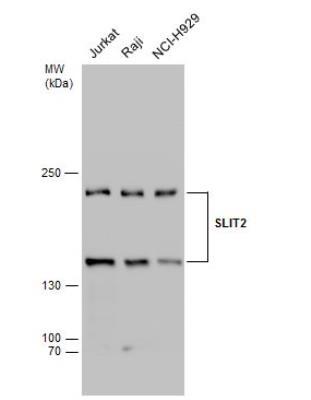
- Western Blot: Slit2 Antibody [NBP2-20398] - Various whole cell extracts (30 ug) were separated by 5% SDS-PAGE, and the membrane was blotted with SLIT2 antibody diluted at 1:1000.
- Submitted by
- Novus Biologicals (provider)
- Main image
- Experimental details
- Western Blot: Slit2 Antibody [NBP2-20398] - Various whole cell extracts (30 ug) were separated by 5% SDS-PAGE, and the membrane was blotted with SLIT2 antibody diluted at 1:1000.
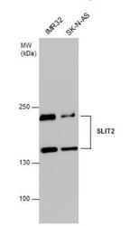
- Submitted by
- Novus Biologicals (provider)
- Main image
- Experimental details
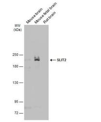
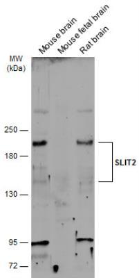
- Western Blot: Slit2 Antibody [NBP2-20398] - Various tissue extracts (30 ug) were separated by 5% SDS-PAGE, and the membrane was blotted with SLIT2 antibody diluted at 1:3000. The HRP-conjugated anti-rabbit IgG antibody (NBP2-19301) was used to detect the primary antibody.
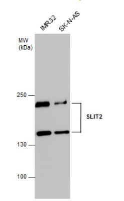
- Submitted by
- Novus Biologicals (provider)
- Main image
- Experimental details
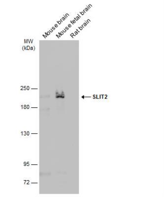
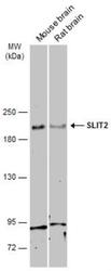
- Western Blot: Slit2 Antibody [NBP2-20398] - Various tissue extracts (30 ug) were separated by 5% SDS-PAGE, and the membrane was blotted with SLIT2 antibody diluted at 1:500. The HRP-conjugated anti-rabbit IgG antibody (NBP2-19301) was used to detect the primary antibody.
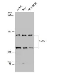
- Submitted by
- Novus Biologicals (provider)
- Main image
- Experimental details
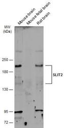
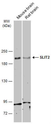
- Western Blot: Slit2 Antibody [NBP2-20398] - Various tissue extracts (50 ug) were separated by 5% SDS-PAGE, and the membrane was blotted with SLIT2 antibody diluted at 1:500. The HRP-conjugated anti-rabbit IgG antibody (NBP2-19301) was used to detect the primary antibody.
Supportive validation
- Submitted by
- Novus Biologicals (provider)
- Main image
- Experimental details
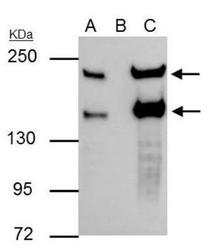
- Immunoprecipitation: Slit2 Antibody [NBP2-20398] - Raji whole cell lysate/extract A : 30 ug whole cell lysate/extract of SLIT2 protein expressing Raji cells B : Control with 3 ug of pre-immune rabbit IgG C : Immunoprecipitation of SLIT2 by 3 ug of SLIT2 antibody 5% SDS-PAGE The immunoprecipitated SLIT2 protein was detected by SLIT2 antibody diluted at 1 : 1000. EasyBlot anti-rabbit IgG (HRP) was used as a secondary reagent.

Supportive validation
- Submitted by
- Novus Biologicals (provider)
- Main image

- Experimental details
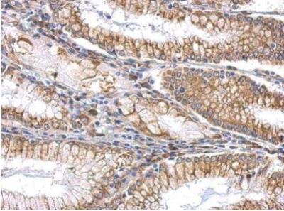
- Immunohistochemistry-Paraffin: Slit2 Antibody [NBP2-20398] - SLIT2 Antibody [NBP2-20398] - Cytosol and membrane on gastric carcinoma .
- Submitted by
- Novus Biologicals (provider)
- Main image
- Experimental details
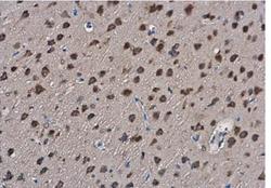
- Immunohistochemistry-Paraffin: Slit2 Antibody [NBP2-20398] - SLIT2 antibody detects SLIT2 protein at cytoplasm in rat brain Paraffin-embedded rat brain diluted at 1:400.

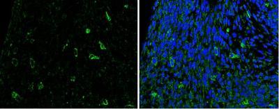
- Submitted by
- Novus Biologicals (provider)
- Main image
- Experimental details
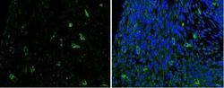
- Immunohistochemistry-Paraffin: Slit2 Antibody [NBP2-20398] - Paraffin-embedded mouse fetal brain. Green: SLIT2 antibody diluted at 1:200. The signal was developed using goat anti-rabbit IgG antibody (Dylight488). Blue: Nuclear staining with Hoechst 33342.