Explore
 Validate
 Learn
Learn Western blot
Antibody data
- Antibody Data
- Antigen structure
- References [3]
- Comments [0]
- Validations
- Western blot [1]
Submit
Validation data
Reference
Comment
Report error
- Product number
- PAB11259 - Provider product page

- Provider
- Abnova Corporation
- Proper citation
- Abnova Corporation Cat#PAB11259, RRID:AB_1714206
- Product name
- SLIT2 polyclonal antibody
- Antibody type
- Polyclonal
- Description
- Rabbit polyclonal antibody raised against synthetic peptide of SLIT2.
- Storage
- Store at 4°C. For long term storage store at -20°C.Aliquot to avoid repeated freezing and thawing.
Submitted references SLIT2-mediated ROBO2 signaling restricts kidney induction to a single site.
Slit protein-mediated inhibition of CXCR4-induced chemotactic and chemoinvasive signaling pathways in breast cancer cells.
Frequent epigenetic inactivation of the SLIT2 gene in gliomas.
Grieshammer U, Le Ma, Plump AS, Wang F, Tessier-Lavigne M, Martin GR
Developmental cell 2004 May;6(5):709-17
Developmental cell 2004 May;6(5):709-17
Slit protein-mediated inhibition of CXCR4-induced chemotactic and chemoinvasive signaling pathways in breast cancer cells.
Prasad A, Fernandis AZ, Rao Y, Ganju RK
The Journal of biological chemistry 2004 Mar 5;279(10):9115-24
The Journal of biological chemistry 2004 Mar 5;279(10):9115-24
Frequent epigenetic inactivation of the SLIT2 gene in gliomas.
Dallol A, Krex D, Hesson L, Eng C, Maher ER, Latif F
Oncogene 2003 Jul 17;22(29):4611-6
Oncogene 2003 Jul 17;22(29):4611-6
No comments: Submit comment
Supportive validation
- Submitted by
- Abnova Corporation (provider)
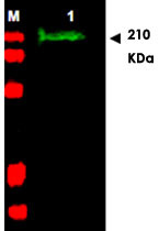
- Main image

- Experimental details
- Western blot using SLIT2 polyclonal antibody (Cat # PAB11259) shows detection of a band at ~165 KDa (Lane 1) corresponding to SLIT2 present in a chicken spinal cord whole cell lysate (arrowhead).Approximately 30 ug of lysate was separated on a 4-20% Tris-Glycine gel by SDS-PAGE and transferred onto nitrocellulose.After blocking the membrane was probed with the primary antibody diluted to 1 : 1,350.Reaction occurred overnight at 4°C followed by washes and reaction with a 1 : 10,000 dilution of IRDye™800 conjugated Gt-a-Rabbit IgG [H&L] MX for 45 min at room temperature (800 nm channel, green).Molecular weight estimation was made by comparison to prestained MW markers in lane M (700 nm channel, red).IRDye™ 800 fluorescence image was captured using the Odyssey® Infrared Imaging System developed by LI-COR.IRDye is a trademark of LI-COR, Inc.