Explore
 Validate
 Learn
Learn Western blot
 Immunohistochemistry
Antibody data
- Antibody Data
- Antigen structure
- References [1]
- Comments [0]
- Validations
- Immunohistochemistry [3]
- Other assay [2]
Submit
Validation data
Reference
Comment
Report error
- Product number
- PA5-31133 - Provider product page

- Provider
- Invitrogen Antibodies
- Product name
- SLIT2 Polyclonal Antibody
- Antibody type
- Polyclonal
- Antigen
- Recombinant full-length protein
- Description
- Recommended positive controls: IMR32, SK-N-AS, mouse brain, rat brain, SLIT2-transfected 293T. Predicted reactivity: Mouse (98%), Rat (95%), Zebrafish (88%), Xenopus laevis (93%), Chicken (97%), Bovine (99%). Store product as a concentrated solution. Centrifuge briefly prior to opening the vial.
- Reactivity
- Human, Mouse, Rat
- Host
- Rabbit
- Isotype
- IgG
- Vial size
- 100 μL
- Concentration
- 0.41 mg/mL
- Storage
- Store at 4°C short term. For long term storage, store at -20°C, avoiding freeze/thaw cycles.
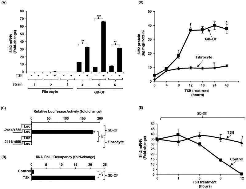
Submitted references Slit2 Modulates the Inflammatory Phenotype of Orbit-Infiltrating Fibrocytes in Graves' Disease.
Fernando R, Grisolia ABD, Lu Y, Atkins S, Smith TJ
Journal of immunology (Baltimore, Md. : 1950) 2018 Jun 15;200(12):3942-3949
Journal of immunology (Baltimore, Md. : 1950) 2018 Jun 15;200(12):3942-3949
No comments: Submit comment
Supportive validation
- Submitted by
- Invitrogen Antibodies (provider)
- Main image
- Experimental details
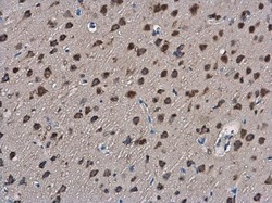
- SLIT2 Polyclonal Antibody detects SLIT2 protein at cytoplasm in rat brain by immunohistochemical analysis. Sample: Paraffin-embedded rat brain. SLIT2 Polyclonal Antibody (Product # PA5-31133) diluted at 1:400. Antigen Retrieval: Citrate buffer, pH 6.0, 15 min.
- Submitted by
- Invitrogen Antibodies (provider)
- Main image
- Experimental details
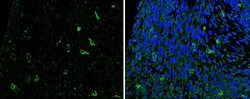
- Immunohistochemistry (Paraffin) analysis of SLIT2 was performed in paraffin-embedded mouse fetal brain tissue using Green: NAGLU Polyclonal Antibody (Product # PA5-28261) at a dilution of 1:200. Blue: Nuclear staining with Hoechst 33342. The signal was developed using a goat anti-rabbit IgG antibody.

- Submitted by
- Invitrogen Antibodies (provider)
- Main image
- Experimental details
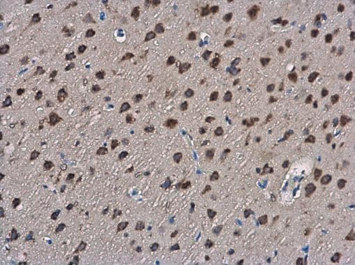
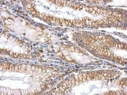
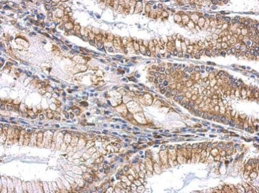
- SLIT2 Polyclonal Antibody detects SLIT2 protein at cytosol and membrane on gastric carcinoma by immunohistochemical analysis. Sample: Paraffin-embedded human gastric carcinoma. SLIT2 Polyclonal Antibody (Product # PA5-31133) dilution: 1:500. Antigen Retrieval: EDTA based buffer, pH 8.0, 15 min.
Supportive validation
- Submitted by
- Invitrogen Antibodies (provider)
- Main image

- Experimental details
- NULL
- Submitted by
- Invitrogen Antibodies (provider)
- Main image

- Experimental details
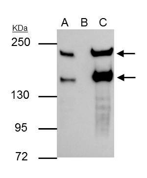
- SLIT2 Polyclonal Antibody immunoprecipitates SLIT2 protein in IP experiments. IP Sample: Raji whole cell lysate/extract A : 30 µg whole cell lysate/extract of SLIT2 protein expressing Raji cells B : Control with 3 µg of pre-immune rabbit IgG C : Immunoprecipitation of SLIT2 by 3 µg of SLIT2 Polyclonal Antibody (Product # PA5-31133) 5% SDS-PAGE The immunoprecipitated SLIT2 protein was detected by SLIT2 Polyclonal Antibody (Product # PA5-31133) diluted at 1:1,000. Anti-rabbit IgG (HRP) was used as a secondary reagent.