 Explore
 Validate
 Learn
LearnHPA041325
antibody from Atlas Antibodies
Targeting: CYP51A1
CP51, CYP51, CYPL1, LDM, P450-14DM, P450L1
 Western blot
 Immunocytochemistry
 Immunohistochemistry
Antibody data
- Antibody Data
- Antigen structure
- References [1]
- Comments [0]
- Validations
- Western blot [2]
- Immunocytochemistry [1]
Submit
Validation data
Reference
Comment
Report error
- Product number
- HPA041325 - Provider product page

- Provider
- Atlas Antibodies
- Proper citation
- Atlas Antibodies Cat#HPA041325, RRID:AB_2677411
- Product name
- Anti-CYP51A1
- Antibody type
- Polyclonal
- Description
- Polyclonal Antibody against Human CYP51A1, Gene description: cytochrome P450, family 51, subfamily A, polypeptide 1, Alternative Gene Names: CP51, CYP51, CYPL1, LDM, P450-14DM, P450L1, Validated applications: ICC, IHC, WB, Uniprot ID: Q16850, Storage: Store at +4°C for short term storage. Long time storage is recommended at -20°C.
- Reactivity
- Human
- Host
- Rabbit
- Conjugate
- Unconjugated
- Isotype
- IgG
- Vial size
- 100 µl
- Concentration
- 0.2 mg/ml
- Storage
- Store at +4°C for short term storage. Long time storage is recommended at -20°C.
- Handling
- The antibody solution should be gently mixed before use.
Submitted references Lung cancer cells survive epidermal growth factor receptor tyrosine kinase inhibitor exposure through upregulation of cholesterol synthesis
Howell M, Green R, Khalil R, Foran E, Quarni W, Nair R, Stevens S, Grinchuk A, Hanna A, Mohapatra S, Mohapatra S
FASEB BioAdvances 2019;2(2):90-105
FASEB BioAdvances 2019;2(2):90-105
No comments: Submit comment
Enhanced validation
Enhanced validation
- Submitted by
- klas2
- Enhanced method
- Genetic validation
- Main image
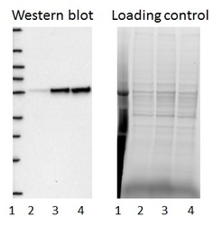
- Experimental details
- Western blot of cell lysate from U-2 OS cells transfected with either siRNA targeting CYP51A1 or control siRNA. Lane 1: Marker (250, 130, 95, 72, 55, 36, 28, 17, 10) Lane 2: Cell lysate from U-2OS cells transfected with siRNA targeting CYP51A1 Lane 3: N/A Lane 4: Cell lysate from U-2OS cells transfected with control siRNA Right image, lane 1-4: loading control
- Sample type
- U-2 OS
- Primary Ab dilution
- 1:205
- Conjugate
- Horseradish Peroxidase
- Secondary Ab
- Secondary Ab
- Secondary Ab dilution
- 1:3000
- Knockdown/Genetic Approaches Application
- Western blot
Enhanced validation
- Submitted by
- Atlas Antibodies (provider)
- Enhanced method
- Genetic validation
- Main image

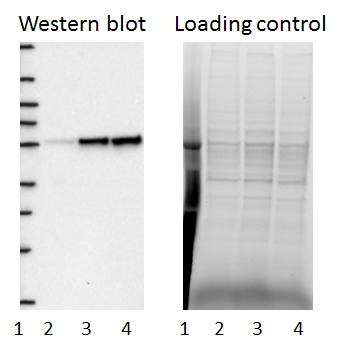
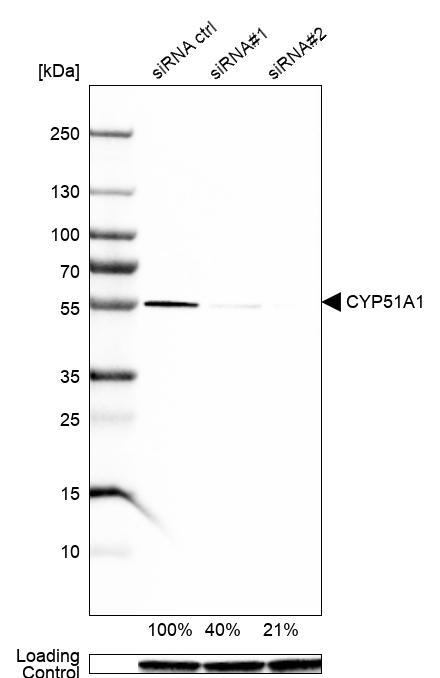
- Experimental details
- Western blot analysis in Caco-2 cells transfected with control siRNA, target specific siRNA probe #1 and #2, using Anti-CYP51A1 antibody. Remaining relative intensity is presented. Loading control: Anti-GAPDH.
- Sample type
- Human
- Protocol
- Protocol
Supportive validation
- Submitted by
- Atlas Antibodies (provider)
- Main image
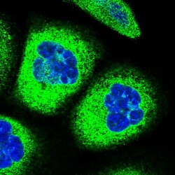
- Experimental details
- Immunofluorescent staining of human cell line A-431 shows localization to endoplasmic reticulum.
- Sample type
- Human
