 Explore
 Validate
 Learn
Learn Immunocytochemistry
Antibody data
- Antibody Data
- Antigen structure
- References [0]
- Comments [0]
- Validations
- Immunocytochemistry [2]
- Immunohistochemistry [1]
Submit
Validation data
Reference
Comment
Report error
- Product number
- MAB3692-100 - Provider product page

- Provider
- R&D Systems
- Product name
- Human Ephrin-A4 Antibody
- Antibody type
- Monoclonal
- Description
- Protein A or G purified from hybridoma culture supernatant. Detects human Ephrin-A4 in direct ELISAs.
- Reactivity
- Human
- Host
- Mouse
- Conjugate
- Unconjugated
- Antigen sequence
P52798- Isotype
- IgG
- Antibody clone number
- 80937
- Vial size
- 100 ug
- Storage
- Use a manual defrost freezer and avoid repeated freeze-thaw cycles. 12 months from date of receipt, -20 to -70 °C as supplied. 1 month, 2 to 8 °C under sterile conditions after reconstitution. 6 months, -20 to -70 °C under sterile conditions after reconstitution.
No comments: Submit comment
Supportive validation
- Submitted by
- R&D Systems (provider)
- Main image

- Experimental details
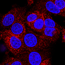
- Ephrin-A4 in MCF-7 Human Cell Line. Ephrin-A4 was detected in immersion fixed MCF-7 human breast cancer cell line using Mouse Anti-Human Ephrin-A4 Monoclonal Antibody (Catalog # MAB3692) at 8 µg/mL for 3 hours at room temperature. Cells were stained using the NorthernLights™ 557-conjugated Anti-Mouse IgG Secondary Antibody (red; Catalog # NL007) and counterstained with DAPI (blue). Specific staining was localized to cytoplasm. View our protocol for Fluorescent ICC Staining of Cells on Coverslips.
- Submitted by
- R&D Systems (provider)
- Main image
- Experimental details
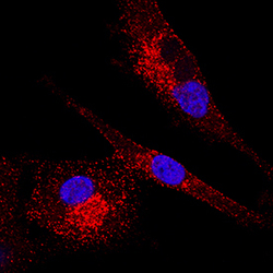
- Ephrin-A4 in U-87 MG Human Cell Line. Ephrin-A4 was detected in immersion fixed U-87 MG human glioblastoma/astrocytoma cell line using Mouse Anti-Human Ephrin-A4 Monoclonal Antibody (Catalog # MAB3692) at 8 µg/mL for 3 hours at room temperature. Cells were stained using the NorthernLights™ 557-conjugated Anti-Mouse IgG Secondary Antibody (red; Catalog # NL007) and counterstained with DAPI (blue). Specific staining was localized to cytoplasm. View our protocol for Fluorescent ICC Staining of Cells on Coverslips.

Supportive validation
- Submitted by
- R&D Systems (provider)
- Main image
- Experimental details
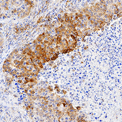
- Ephrin-A4 in Human Squamous Cell Carcinoma. Ephrin-A4 was detected in immersion fixed paraffin-embedded sections of human squamous cell carcinoma using Mouse Anti-Human Ephrin-A4 Monoclonal Antibody (Catalog # MAB3692) at 5 µg/mL for 1 hour at room temperature followed by incubation with the Anti-Mouse IgG VisUCyte™ HRP Polymer Antibody (Catalog # VC001). Tissue was stained using DAB (brown) and counterstained with hematoxylin (blue). Specific staining was localized to cytoplasm in cancer cells. View our protocol for IHC Staining with VisUCyte HRP Polymer Detection Reagents.
