 Explore
 Validate
 Learn
Learn Western blot
 Immunocytochemistry
Antibody data
- Antibody Data
- Antigen structure
- References [2]
- Comments [0]
- Validations
- Immunocytochemistry [1]
- Immunohistochemistry [1]
Submit
Validation data
Reference
Comment
Report error
- Product number
- AF369 - Provider product page

- Provider
- R&D Systems
- Product name
- Human Ephrin-A4 Antibody
- Antibody type
- Polyclonal
- Description
- Antigen Affinity-purified. Detects human Ephrin-A4 in direct ELISAs and Western blots. In direct ELISAs, less than 25% cross-reactivity with recombinant mouse Ephrin-A4 is observed.
- Reactivity
- Human
- Host
- Goat
- Conjugate
- Unconjugated
- Antigen sequence
P52798- Isotype
- IgG
- Vial size
- 100 ug
- Concentration
- LYOPH
- Storage
- Use a manual defrost freezer and avoid repeated freeze-thaw cycles. 12 months from date of receipt, -20 to -70 °C as supplied. 1 month, 2 to 8 °C under sterile conditions after reconstitution. 6 months, -20 to -70 °C under sterile conditions after reconstitution.
Submitted references Identification of microRNA signature in the progression of gestational trophoblastic disease.
Signaling through ephrin-A ligand leads to activation of Src-family kinases, Akt phosphorylation, and inhibition of antigen receptor-induced apoptosis.
Zhao JR, Cheng WW, Wang YX, Cai M, Wu WB, Zhang HJ
Cell death & disease 2018 Jan 24;9(2):94
Cell death & disease 2018 Jan 24;9(2):94
Signaling through ephrin-A ligand leads to activation of Src-family kinases, Akt phosphorylation, and inhibition of antigen receptor-induced apoptosis.
Holen HL, Shadidi M, Narvhus K, Kjøsnes O, Tierens A, Aasheim HC
Journal of leukocyte biology 2008 Oct;84(4):1183-91
Journal of leukocyte biology 2008 Oct;84(4):1183-91
No comments: Submit comment
Supportive validation
- Submitted by
- R&D Systems (provider)
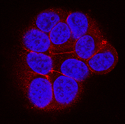
- Main image
- Experimental details
- Ephrin-A4 in MCF-7 Human Cell Line. Ephrin-A4 was detected in immersion fixed MCF-7 human breast cancer cell line using Goat Anti-Human Ephrin-A4 Antigen Affinity-purified Polyclonal Antibody (Catalog # AF369) at 5 µg/mL for 3 hours at room temperature. Cells were stained using the NorthernLights™ 557-conjugated Anti-Goat IgG Secondary Antibody (red; Catalog # NL001) and counterstained with DAPI (blue). Specific staining was localized to plasma membrane. View our protocol for Fluorescent ICC Staining of Cells on Coverslips.

Supportive validation
- Submitted by
- R&D Systems (provider)
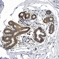
- Main image
- Experimental details
- Ephrin-A4 in Human Mammary Glands. Ephrin-A4 was detected in immersion fixed paraffin-embedded sections of normal human mammary glands using Goat Anti-Human Ephrin-A4 Antigen Affinity-purified Polyclonal Antibody (Catalog # AF369) at 3 µg/mL for 1 hour at room temperature followed by incubation with the Anti-Goat IgG VisUCyte™ HRP Polymer Antibody (Catalog # VC004). Tissue was stained using DAB (brown) and counterstained with hematoxylin (blue). Specific staining was localized to cytoplasm and plasma membrane of glandular epithelium. View our protocol for IHC Staining with VisUCyte HRP Polymer Detection Reagents.
