 Explore
 Validate
 Learn
Learn Western blot
 Immunohistochemistry
Antibody data
- Antibody Data
- Antigen structure
- References [6]
- Comments [0]
- Validations
- Western blot [2]
- Immunocytochemistry [2]
Submit
Validation data
Reference
Comment
Report error
- Product number
- HPA000525 - Provider product page

- Provider
- Atlas Antibodies
- Proper citation
- Atlas Antibodies Cat#HPA000525, RRID:AB_1078106
- Product name
- Anti-ADSL
- Antibody type
- Polyclonal
- Description
- Polyclonal Antibody against Human ADSL, Gene description: adenylosuccinate lyase, Validated applications: ICC, IHC, WB, Uniprot ID: P30566, Storage: Store at +4°C for short term storage. Long time storage is recommended at -20°C.
- Reactivity
- Human
- Host
- Rabbit
- Conjugate
- Unconjugated
- Isotype
- IgG
- Vial size
- 100 µl
- Concentration
- 0.1 mg/ml
- Storage
- Store at +4°C for short term storage. Long time storage is recommended at -20°C.
- Handling
- The antibody solution should be gently mixed before use.
Submitted references MicroRNA-21 guide and passenger strand regulation of adenylosuccinate lyase-mediated purine metabolism promotes transition to an EGFR-TKI-tolerant persister state
Pathway-specific effects of ADSL deficiency on neurodevelopment
Prolyl hydroxylase substrate adenylosuccinate lyase is an oncogenic driver in triple negative breast cancer
Metabolomic profiling of human lung tumor tissues – nucleotide metabolism as a candidate for therapeutic interventions and biomarkers
Systematic validation of antibody binding and protein subcellular localization using siRNA and confocal microscopy
Mutations of ATIC and ADSL affect purinosome assembly in cultured skin fibroblasts from patients with AICA-ribosiduria and ADSL deficiency
Zhang W, Skiados N, Aftab F, Moreno C, Silva L, Corbilla P, Asara J, Hata A, Slack F
Cancer Gene Therapy 2022;29(12):1878-1894
Cancer Gene Therapy 2022;29(12):1878-1894
Pathway-specific effects of ADSL deficiency on neurodevelopment
Gerhards J, Dutto I, Herrera A, Souckova O, Škopová V, Smak J, Junza A, Yanes O, Boeckx C, Burkhalter M, Zikánová M, Pons S, Philipp M, Lüders J, Stracker T
eLife 2022;11
eLife 2022;11
Prolyl hydroxylase substrate adenylosuccinate lyase is an oncogenic driver in triple negative breast cancer
Zurlo G, Liu X, Takada M, Fan C, Simon J, Ptacek T, Rodriguez J, von Kriegsheim A, Liu J, Locasale J, Robinson A, Zhang J, Holler J, Kim B, Zikánová M, Bierau J, Xie L, Chen X, Li M, Perou C, Zhang Q
Nature Communications 2019;10(1)
Nature Communications 2019;10(1)
Metabolomic profiling of human lung tumor tissues – nucleotide metabolism as a candidate for therapeutic interventions and biomarkers
Moreno P, Jiménez‐Jiménez C, Garrido‐Rodríguez M, Calderón‐Santiago M, Molina S, Lara‐Chica M, Priego‐Capote F, Salvatierra Á, Muñoz E, Calzado M
Molecular Oncology 2018;12(10):1778-1796
Molecular Oncology 2018;12(10):1778-1796
Systematic validation of antibody binding and protein subcellular localization using siRNA and confocal microscopy
Stadler C, Hjelmare M, Neumann B, Jonasson K, Pepperkok R, Uhlén M, Lundberg E
Journal of Proteomics 2012;75(7):2236-2251
Journal of Proteomics 2012;75(7):2236-2251
Mutations of ATIC and ADSL affect purinosome assembly in cultured skin fibroblasts from patients with AICA-ribosiduria and ADSL deficiency
Baresova V, Skopova V, Sikora J, Patterson D, Sovova J, Zikanova M, Kmoch S
Human Molecular Genetics 2011;21(7):1534-1543
Human Molecular Genetics 2011;21(7):1534-1543
No comments: Submit comment
Enhanced validation
Enhanced validation
- Submitted by
- klas2
- Enhanced method
- Genetic validation
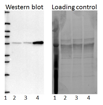
- Main image
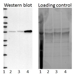
- Experimental details
- Western blot of cell lysate from U-2 OS cells transfected with either siRNA targeting ADSL or control siRNA. Lane 1: Marker (250, 130, 95, 72, 55, 36, 28, 17, 10) Lane 2: Cell lysate from U-2OS cells transfected with siRNA targeting ADSL Lane 3: N/A Lane 4: Cell lysate from U-2OS cells transfected with control siRNA Right image, lane 1-4: loading control
- Sample type
- U-2 OS
- Primary Ab dilution
- 1:119
- Conjugate
- Horseradish Peroxidase
- Secondary Ab
- Secondary Ab
- Secondary Ab dilution
- 1:3000
- Knockdown/Genetic Approaches Application
- Western blot
Enhanced validation
- Submitted by
- Atlas Antibodies (provider)
- Enhanced method
- Genetic validation
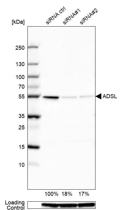
- Main image

- Experimental details
- Western blot analysis in U2OS cells transfected with control siRNA, target specific siRNA probe #1 and #2, using Anti-ADSL antibody. Remaining relative intensity is presented. Loading control: Anti-GAPDH.
- Sample type
- Human
- Protocol
- Protocol
Enhanced validation
Supportive validation
- Submitted by
- 55af80e3e0991
- Enhanced method
- Genetic validation
- Main image
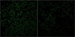
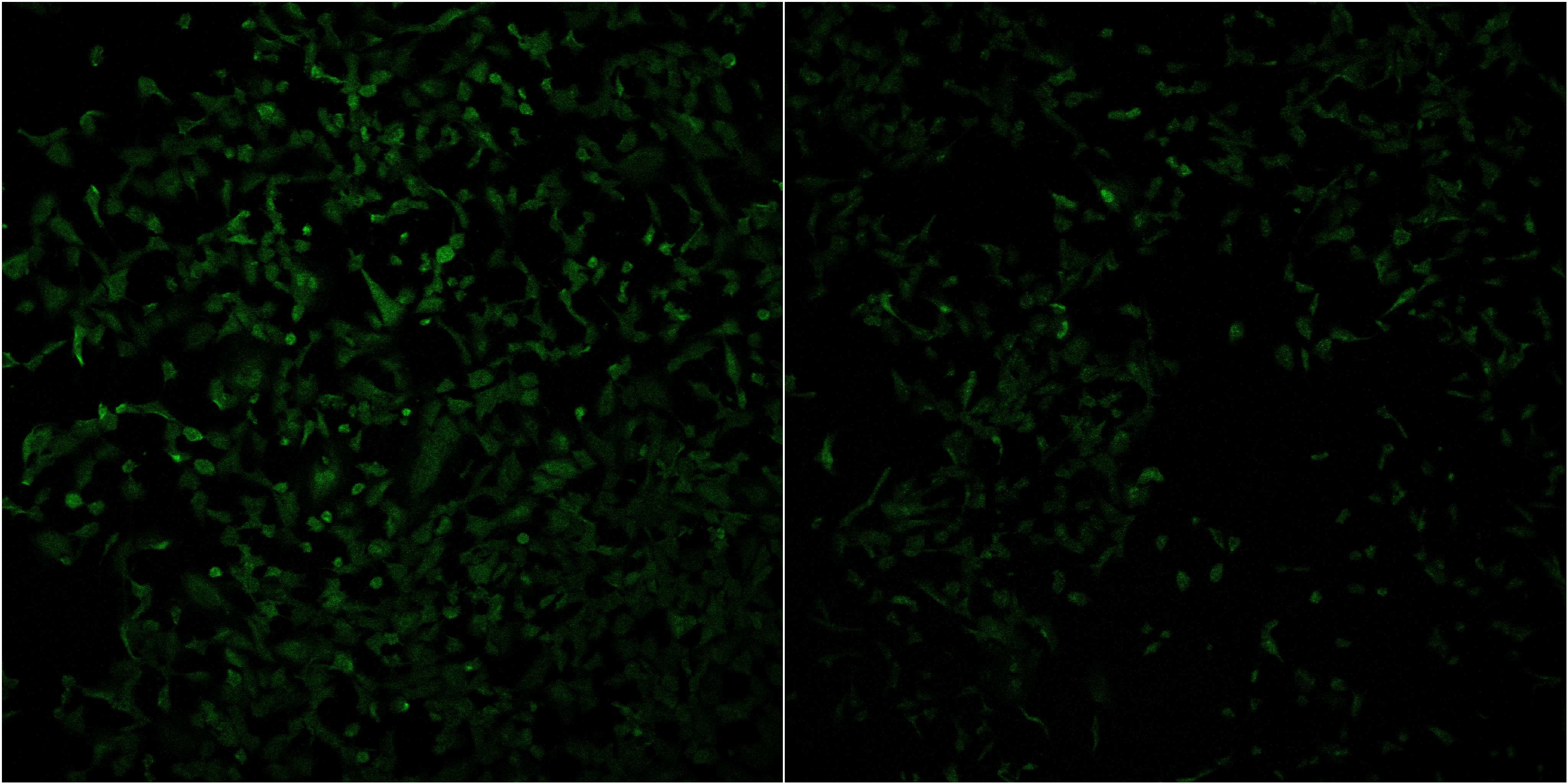
- Experimental details
- Confocal images of immunofluorescently stained human U-2 OS cells.The protein ADSL is shown in green. The image to the left show cells transfected with control siRNA and the image to the right show cells where ADSL has been downregulated with specific siRNA.
- Sample type
- U-2 OS cells
- Primary Ab dilution
- 1:53
- Secondary Ab
- Secondary Ab
- Secondary Ab dilution
- 1:800
- Knockdown/Genetic Approaches Application
- Immunocytochemistry
Supportive validation
- Submitted by
- Atlas Antibodies (provider)
- Main image
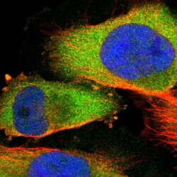
- Experimental details
- Immunofluorescent staining of human cell line U-251 MG shows localization to cytosol.
- Sample type
- Human
