Explore
 Validate
 Learn
Learn Western blot
 Immunocytochemistry
Antibody data
- Antibody Data
- Antigen structure
- References [2]
- Comments [0]
- Validations
- Western blot [2]
- Immunohistochemistry [8]
Submit
Validation data
Reference
Comment
Report error
- Product number
- NBP1-87406 - Provider product page

- Provider
- Novus Biologicals
- Proper citation
- Novus Cat#NBP1-87406, RRID:AB_11021353
- Product name
- Rabbit Polyclonal Adenylosuccinate Lyase Antibody
- Antibody type
- Polyclonal
- Description
- Immunogen affinity purified. Specificity of human Adenylosuccinate Lyase antibody verified on a Protein Array containing target protein plus 383 other non-specific proteins.
- Reactivity
- Human
- Host
- Rabbit
- Isotype
- IgG
- Vial size
- 0.1 ml
- Storage
- Store at 4C short term. Aliquot and store at -20C long term. Avoid freeze-thaw cycles.
Submitted references Systematic validation of antibody binding and protein subcellular localization using siRNA and confocal microscopy.
Mutations of ATIC and ADSL affect purinosome assembly in cultured skin fibroblasts from patients with AICA-ribosiduria and ADSL deficiency.
Stadler C, Hjelmare M, Neumann B, Jonasson K, Pepperkok R, Uhlén M, Lundberg E
Journal of proteomics 2012 Apr 3;75(7):2236-51
Journal of proteomics 2012 Apr 3;75(7):2236-51
Mutations of ATIC and ADSL affect purinosome assembly in cultured skin fibroblasts from patients with AICA-ribosiduria and ADSL deficiency.
Baresova V, Skopova V, Sikora J, Patterson D, Sovova J, Zikanova M, Kmoch S
Human molecular genetics 2012 Apr 1;21(7):1534-43
Human molecular genetics 2012 Apr 1;21(7):1534-43
No comments: Submit comment
Supportive validation
- Submitted by
- Novus Biologicals (provider)
- Main image
- Experimental details
- Western Blot: Adenylosuccinate Lyase Antibody [NBP1-87406] - Analysis in human cell line HL-60.

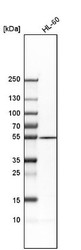
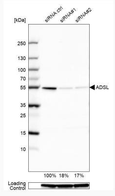
- Submitted by
- Novus Biologicals (provider)
- Main image

- Experimental details
- Western Blot: Adenylosuccinate Lyase Antibody [NBP1-87406] - Analysis in U2OS cells transfected with control siRNA, target specific siRNA probe #1 and #2, using Anti-ADSL antibody. Remaining relative intensity is presented. Loading control: Anti-GAPDH.
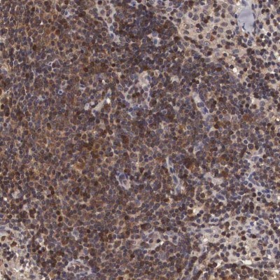
Supportive validation
- Submitted by
- Novus Biologicals (provider)
- Main image

- Experimental details
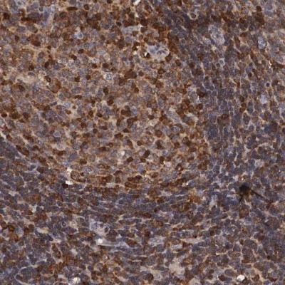
- Immunohistochemistry-Paraffin: Adenylosuccinate Lyase Antibody [NBP1-87406] - Staining of human lymph node shows high expression.
- Submitted by
- Novus Biologicals (provider)
- Main image
- Experimental details
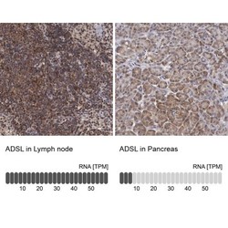
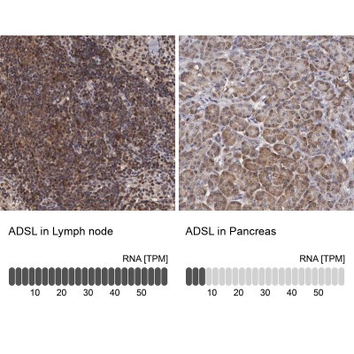
- Immunohistochemistry-Paraffin: Adenylosuccinate Lyase Antibody [NBP1-87406] - Staining in human lymph node and pancreas tissues using anti-ADSL antibody. Corresponding ADSL RNA-seq data are presented for the same tissues.
- Submitted by
- Novus Biologicals (provider)
- Main image
- Experimental details
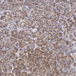
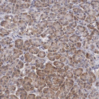
- Immunohistochemistry-Paraffin: Adenylosuccinate Lyase Antibody [NBP1-87406] - Staining of human pancreas shows low expression as expected.
- Submitted by
- Novus Biologicals (provider)
- Main image
- Experimental details
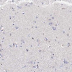
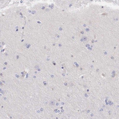
- Immunohistochemistry-Paraffin: Adenylosuccinate Lyase Antibody [NBP1-87406] - Staining of human cerebral cortex shows no positivity in neuronal cells.
- Submitted by
- Novus Biologicals (provider)
- Main image
- Experimental details
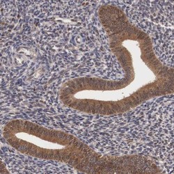
- Immunohistochemistry-Paraffin: Adenylosuccinate Lyase Antibody [NBP1-87406] - Staining of human endometrium shows moderate cytoplasmic positivity in glandular cells.

- Submitted by
- Novus Biologicals (provider)
- Main image

- Experimental details
- Immunohistochemistry-Paraffin: Adenylosuccinate Lyase Antibody [NBP1-87406] - Staining of human lymphoid tissues shows moderate cytoplasmic positivity in germinal center cells.
- Submitted by
- Novus Biologicals (provider)
- Main image

- Experimental details
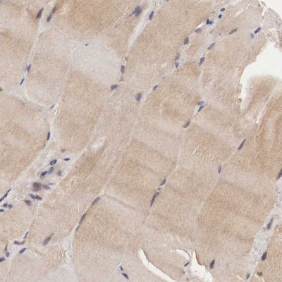
- Immunohistochemistry-Paraffin: Adenylosuccinate Lyase Antibody [NBP1-87406] - Staining of human skeletal muscle shows moderate cytoplasmic positivity in myocytes.
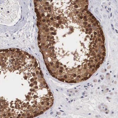
- Submitted by
- Novus Biologicals (provider)
- Main image

- Experimental details
- Immunohistochemistry-Paraffin: Adenylosuccinate Lyase Antibody [NBP1-87406] - Staining of human testis shows moderate cytoplasmic positivity in cells in seminiferous ducts.