Explore
 Validate
 Learn
Learn Western blot
 Immunocytochemistry
Antibody data
- Antibody Data
- Antigen structure
- References [0]
- Comments [0]
- Validations
- Immunocytochemistry [3]
- Immunohistochemistry [1]
- Chromatin Immunoprecipitation [2]
Submit
Validation data
Reference
Comment
Report error
- Product number
- PA1-9005 - Provider product page

- Provider
- Invitrogen Antibodies
- Product name
- Androgen Receptor Polyclonal Antibody
- Antibody type
- Polyclonal
- Antigen
- Synthetic peptide
- Description
- This antibody is predicted to react with bovine, canine, mouse, porcine and rat based on sequence homology. This antibody is tested in Peptide ELISA: antibody detection limit dilution 64,000.
- Reactivity
- Human
- Host
- Goat
- Isotype
- IgG
- Vial size
- 100 μg
- Concentration
- 0.5 mg/mL
- Storage
- -20°C, Avoid Freeze/Thaw Cycles
No comments: Submit comment
Supportive validation
- Submitted by
- Invitrogen Antibodies (provider)
- Main image

- Experimental details
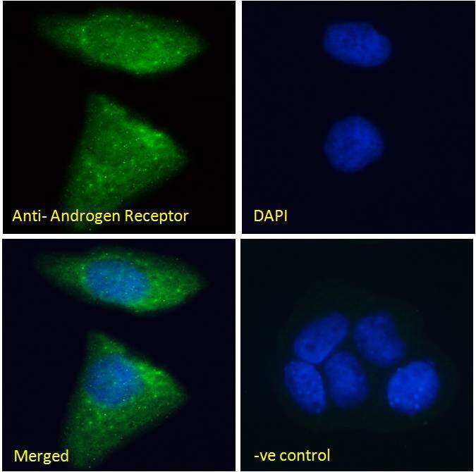
- Immunocytochemistry analysis of Androgen Receptor using Androgen Receptor Polyclonal Antibody (Product # PA1-9005) in paraformaldehyde fixed MCF7 cells, permeabilized with 0.15% Triton. Primary incubation 1hr (10 µg/mL) followed by Alexa Fluor 488 secondary antibody (2 µg/mL), showing Mitochondrial/cytoplasmic staining. The nuclear stain is DAPI (blue). Negative control: Unimmunized goat IgG (10 µg/mL) followed by Alexa Fluor 488 secondary antibody (2 µg/mL).
- Submitted by
- Invitrogen Antibodies (provider)
- Main image

- Experimental details
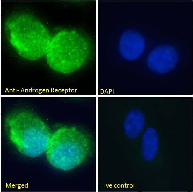
- Immunofluorescence analysis of Androgen Receptor in U2OS cells using a Androgen Receptor monoclonal antibody (Product # PA1-9005) at 10 µg/mL for1hr. The cells were paraformaldehyde fixed and permeabilized with 0.15% Triton. Primary incubation was followed by Alexa Fluor 488 secondary antibody (2 µg/mL) showing Mitochondrial/cytoplasmic staining. The nuclear stain is DAPI (blue). Negative control: Unimmunized goat IgG (10 µg/mL)followed by Alexa Fluor 488 secondary antibody (2 µg/mL).
- Submitted by
- Invitrogen Antibodies (provider)
- Main image

- Experimental details
- Immunocytochemistry analysis of Androgen Receptor using Androgen Receptor Polyclonal Antibody (Product # PA1-9005) in paraformaldehyde fixed U2OS cells, permeabilized with 0.15% Triton. Primary incubation 1hr (10 µg/mL) followed by Alexa Fluor 488 secondary antibody (2 µg/mL), showing Mitochondrial/cytoplasmic staining. The nuclear stain is DAPI (blue). Negative control: Unimmunized goat IgG (10 µg/mL) followed by Alexa Fluor 488 secondary antibody (2 µg/mL).

Supportive validation
- Submitted by
- Invitrogen Antibodies (provider)
- Main image
- Experimental details
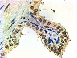
- Immunohistochemical analysis of Androgen Receptor in paraffin embedded human prostate using a Androgen Receptor polyclonal antibody (Product #PA1-9005) at a concentration of 2 µg/mL. Steamed antigen retrieval was performed with pH 6 buffered citrate. Samples were then stained with alkaline phosphatase.

Supportive validation
- Submitted by
- Invitrogen Antibodies (provider)
- Main image

- Experimental details
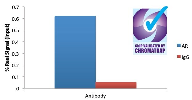
- ChIP analysis of Androgen Receptor in 1 µg of DHT-treated HEC50 chromatin using 2 µg of an Androgen Receptor Polyclonal Antibody (Product #PA1-9005). FKBP5 enrichment was measured.

- Submitted by
- Invitrogen Antibodies (provider)
- Main image
- Experimental details
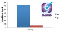
- ChIP assay of 2 µg Androgen Receptor (Product # PA1-9005) with 1 µg DHT-treated HEC50 chromatin using the Chromatrap spin column sonication kit (Protein G) measuring FKBP5 enrichment.