 Explore
 Validate
 Learn
LearnPA5-50656
antibody from Invitrogen Antibodies
Targeting: EPPIN
CT71, dJ461P17.2, EPPIN1, EPPIN2, EPPIN3, SPINLW1, WAP7, WFDC7
 Western blot
Antibody data
- Antibody Data
- Antigen structure
- References [0]
- Comments [0]
- Validations
- Western blot [3]
- Immunohistochemistry [1]
Submit
Validation data
Reference
Comment
Report error
- Product number
- PA5-50656 - Provider product page

- Provider
- Invitrogen Antibodies
- Product name
- SPINLW1 Polyclonal Antibody
- Antibody type
- Polyclonal
- Antigen
- Synthetic peptide
- Description
- The antibody detects endogenous levels of total EPPIN protein.
- Reactivity
- Human, Mouse
- Host
- Rabbit
- Isotype
- IgG
- Vial size
- 100 µL
- Concentration
- 2.4 mg/mL
- Storage
- -20°C
No comments: Submit comment
Supportive validation
- Submitted by
- Invitrogen Antibodies (provider)
- Main image

- Experimental details
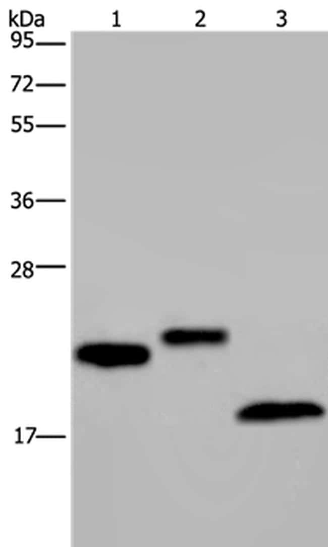
- Western blot analysis of EPPIN was performed by loading (from left to right): NIH/3T3 cell, human testis and mouse fat tissue lysates (40µg) on to a 10% SDS-PAGE gel. Proteins were transferred to a membrane and the membrane was probed with a EPPIN antibody (Product # PA5-50656) at a 1/400 dilution for 20 seconds, followed by a secondary antibody at 1/8000.

- Submitted by
- Invitrogen Antibodies (provider)
- Main image

- Experimental details
- Western blot analysis of EPPIN was performed by loading (from left to right): NIH/3T3 cell, human testis and mouse fat tissue lysates (40µg) on to a 10% SDS-PAGE gel. Proteins were transferred to a membrane and the membrane was probed with a EPPIN antibody (Product # PA5-50656) at a 1/400 dilution for 20 seconds, followed by a secondary antibody at 1/8000.
Supportive validation
- Submitted by
- Invitrogen Antibodies (provider)
- Main image
- Experimental details
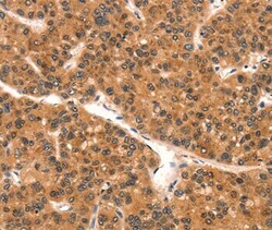
- Immunohistochemical analysis of SPINLW1 in paraffin embedded Human liver cancer tissue using SPINLW1 Polyclonal Antibody (Product # PA5-50656) at a 1:25 dilution.
