 Explore
 Validate
 Learn
Learn Western blot
Antibody data
- Antibody Data
- Antigen structure
- References [1]
- Comments [0]
- Validations
- Western blot [1]
- Immunocytochemistry [1]
- Immunohistochemistry [1]
Submit
Validation data
Reference
Comment
Report error
- Product number
- PA5-36742 - Provider product page

- Provider
- Invitrogen Antibodies
- Product name
- Phospho-Bcl-2 (Thr69) Polyclonal Antibody
- Antibody type
- Polyclonal
- Antigen
- Synthetic peptide
- Description
- This antibody detects endogenous protein at a molecular weight of 26 kDa. Purity is >95% by SDS-PAGE.
- Reactivity
- Human, Mouse, Rat
- Host
- Rabbit
- Isotype
- IgG
- Vial size
- 100 µL
- Concentration
- 1 mg/mL
- Storage
- Store at 4°C short term. For long term storage, store at -20°C, avoiding freeze/thaw cycles.
Submitted references Serine-70 phosphorylated Bcl-2 prevents oxidative stress-induced DNA damage by modulating the mitochondrial redox metabolism.
Chong SJF, Iskandar K, Lai JXH, Qu J, Raman D, Valentin R, Herbaux C, Collins M, Low ICC, Loh T, Davids M, Pervaiz S
Nucleic acids research 2020 Dec 16;48(22):12727-12745
Nucleic acids research 2020 Dec 16;48(22):12727-12745
No comments: Submit comment
Supportive validation
- Submitted by
- Invitrogen Antibodies (provider)
- Main image

- Experimental details
- Western blot analysis of Phospho-Bcl-2 pThr69 using Phospho-Bcl-2 pThr69 polyclonal antibody (Product # PA5-36742) at a dilution of 1:500. Lane 1: HepG2 cell lysate treated with UV, Lane 2: NIH-3T3 cell lysate treated with H2O2, Lane 3: PC12 cell lysate treated with H2O2.

Supportive validation
- Submitted by
- Invitrogen Antibodies (provider)
- Main image

- Experimental details
- Immunofluorescence analysis of Phospho-Bcl-2 (Thr69) was performed using 70% confluent log phase A-431 cells treated with 1mM of Paclitaxel for 20 hours. The cells were fixed with 4% paraformaldehyde for 10 minutes, permeabilized with 0.1% Triton™ X-100 for 15 minutes, and blocked with 1% BSA for 1 hour at room temperature. The cells were labeled with Phospho-Bcl-2 (Thr69) Rabbit Polyclonal Antibody (Product # PA5-36742) at 5 microgram/mL in 0.1% BSA, incubated at 4 degree Celsius overnight and then labeled with Goat anti-Rabbit IgG (H+L) Superclonal™ Secondary Antibody, Alexa Fluor® 488 conjugate (Product # A27034) at a dilution of 1:2000 for 45 minutes at room temperature (Panel a: green). Nuclei (Panel b: blue) were stained with SlowFade® Gold Antifade Mountant with DAPI (Product # S36938). F-actin (Panel c: red) was stained with Rhodamine Phalloidin (Product # R415, 1:300). Panel d represents the merged image showing cytoplasmic localization. Panel e shows untreated cells with reduced signal. Panel f represents control cells with no primary antibody to assess background. The images were captured at 60X magnification.

Supportive validation
- Submitted by
- Invitrogen Antibodies (provider)
- Main image
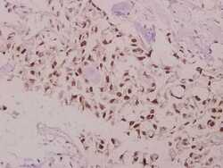
- Experimental details
- Immunohistochemical analysis of Phospho-Bcl-2 pThr69 in paraffin-embedded human colorectal carcinoma using Phospho-Bcl-2 pThr69 polyclonal antibody (Product # PA5-36742) at a dilution of 1:50.
