 Explore
 Validate
 Learn
Learn Western blot
 Flow cytometry
Antibody data
- Antibody Data
- Antigen structure
- References [13]
- Comments [0]
- Validations
- Western blot [2]
- Immunocytochemistry [1]
- Immunohistochemistry [1]
Submit
Validation data
Reference
Comment
Report error
- Product number
- MA5-13666 - Provider product page

- Provider
- Invitrogen Antibodies
- Product name
- Anti-Bcl-2 Monoclonal Antibody (8C8)
- Antibody type
- Monoclonal
- Antigen
- Synthetic peptide
- Description
- MA5-13666 targets BCL-2 alpha in FACS, IF, IHC (P), and WB applications and shows reactivity with Human and Non-human primate samples. The MA5-13666 immunogen is a Synthetic peptide, aa 41-54 (Cys-GAAPAPGIFSSQPG) of human bcl-2 protein.
- Reactivity
- Human
- Host
- Mouse
- Isotype
- IgG
- Antibody clone number
- 8C8
- Vial size
- 500 µL
- Concentration
- 0.2 mg/ml
- Storage
- 4° C
Submitted references Estrogen Receptor-β Up-Regulates IGF1R Expression and Activity to Inhibit Apoptosis and Increase Growth of Medulloblastoma.
Effects of estrogen replacement therapy on apoptosis and vascular endothelial growth factor expression in ocular surface epithelial cells: An experimental study.
Immunohistochemistry with apoptotic-antiapoptotic proteins (p53, p21, bax, bcl-2), c-kit, telomerase, and metallothionein as a diagnostic aid in benign, borderline, and malignant serous and mucinous ovarian tumors.
Clinicopathological significance of cathepsin D expression in non-small cell lung cancer is conditional on apoptosis-associated protein phenotype: an immunohistochemistry study.
The expression of IgM is helpful in the differentiation of primary cutaneous diffuse large B cell lymphoma and follicle center lymphoma.
Recurrence of benign meningiomas: predictive value of proliferative index, BCL2, p53, hormonal receptors and HER2 expression.
Radiotherapy in laryngeal carcinoma: can a panel of 13 markers predict response?
Prognostic significance of cell proliferation and apoptosis-regulating proteins in Epstein-Barr virus positive and negative pediatric Hodgkin lymphoma.
Curcumin-induced apoptosis in human leukemia cell HL-60 is associated with inhibition of telomerase activity.
Incidental calcifying fibrous tumor of the stomach presenting as a polyp.
Tea-induced apoptosis in human leukemia K562 cells as assessed by comet formation.
Induction of apoptosis in human leukemia cells by black tea and its polyphenol theaflavin.
Prognostic factors in transitional cell cancer of the bladder: an emerging role for Bcl-2 and p53.
Cookman CJ, Belcher SM
Endocrinology 2015 Jul;156(7):2395-408
Endocrinology 2015 Jul;156(7):2395-408
Effects of estrogen replacement therapy on apoptosis and vascular endothelial growth factor expression in ocular surface epithelial cells: An experimental study.
Ozcura F, Dündar SO, Cetin ED, Beder N, Dündar M
International journal of ophthalmology 2012;5(1):64-8
International journal of ophthalmology 2012;5(1):64-8
Immunohistochemistry with apoptotic-antiapoptotic proteins (p53, p21, bax, bcl-2), c-kit, telomerase, and metallothionein as a diagnostic aid in benign, borderline, and malignant serous and mucinous ovarian tumors.
Ozer H, Yenicesu G, Arici S, Cetin M, Tuncer E, Cetin A
Diagnostic pathology 2012 Sep 20;7:124
Diagnostic pathology 2012 Sep 20;7:124
Clinicopathological significance of cathepsin D expression in non-small cell lung cancer is conditional on apoptosis-associated protein phenotype: an immunohistochemistry study.
Fan C, Lin X, Wang E
Tumour biology : the journal of the International Society for Oncodevelopmental Biology and Medicine 2012 Aug;33(4):1045-52
Tumour biology : the journal of the International Society for Oncodevelopmental Biology and Medicine 2012 Aug;33(4):1045-52
The expression of IgM is helpful in the differentiation of primary cutaneous diffuse large B cell lymphoma and follicle center lymphoma.
Demirkesen C, Tüzüner N, Esen T, Lebe B, Ozkal S
Leukemia research 2011 Sep;35(9):1269-72
Leukemia research 2011 Sep;35(9):1269-72
Recurrence of benign meningiomas: predictive value of proliferative index, BCL2, p53, hormonal receptors and HER2 expression.
Abdelzaher E, El-Gendi SM, Yehya A, Gowil AG
British journal of neurosurgery 2011 Dec;25(6):707-13
British journal of neurosurgery 2011 Dec;25(6):707-13
Radiotherapy in laryngeal carcinoma: can a panel of 13 markers predict response?
Wildeman MA, Gibcus JH, Hauptmann M, Begg AC, van Velthuysen ML, Hoebers FJ, Mastik MF, Schuuring E, van der Wal JE, van den Brekel MW
The Laryngoscope 2009 Feb;119(2):316-22
The Laryngoscope 2009 Feb;119(2):316-22
Prognostic significance of cell proliferation and apoptosis-regulating proteins in Epstein-Barr virus positive and negative pediatric Hodgkin lymphoma.
Aktaş S, Kargi A, Olgun N, Diniz G, Erbay A, Vergin C
Lymphatic research and biology 2007;5(3):175-82
Lymphatic research and biology 2007;5(3):175-82
Curcumin-induced apoptosis in human leukemia cell HL-60 is associated with inhibition of telomerase activity.
Mukherjee Nee Chakraborty S, Ghosh U, Bhattacharyya NP, Bhattacharya RK, Dey S, Roy M
Molecular and cellular biochemistry 2007 Mar;297(1-2):31-9
Molecular and cellular biochemistry 2007 Mar;297(1-2):31-9
Incidental calcifying fibrous tumor of the stomach presenting as a polyp.
Elpek GO, Küpesiz GY, Ogüs M
Pathology international 2006 Apr;56(4):227-31
Pathology international 2006 Apr;56(4):227-31
Tea-induced apoptosis in human leukemia K562 cells as assessed by comet formation.
Chakraborty S, Kundu T, Dey S, Bhattacharya RK, Siddiqi M, Roy M
Asian Pacific journal of cancer prevention : APJCP 2006 Apr-Jun;7(2):201-7
Asian Pacific journal of cancer prevention : APJCP 2006 Apr-Jun;7(2):201-7
Induction of apoptosis in human leukemia cells by black tea and its polyphenol theaflavin.
Kundu T, Dey S, Roy M, Siddiqi M, Bhattacharya RK
Cancer letters 2005 Dec 8;230(1):111-21
Cancer letters 2005 Dec 8;230(1):111-21
Prognostic factors in transitional cell cancer of the bladder: an emerging role for Bcl-2 and p53.
Ong F, Moonen LM, Gallee MP, ten Bosch C, Zerp SF, Hart AA, Bartelink H, Verheij M
Radiotherapy and oncology : journal of the European Society for Therapeutic Radiology and Oncology 2001 Nov;61(2):169-75
Radiotherapy and oncology : journal of the European Society for Therapeutic Radiology and Oncology 2001 Nov;61(2):169-75
No comments: Submit comment
Supportive validation
- Submitted by
- Invitrogen Antibodies (provider)
- Main image
- Experimental details
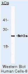
- Western blot of BCL-2 alpha using BCL-2 alpha Monoclonal Antibody (Product # MA5-13666) on HeLa Cells.

- Submitted by
- Invitrogen Antibodies (provider)
- Main image

- Experimental details
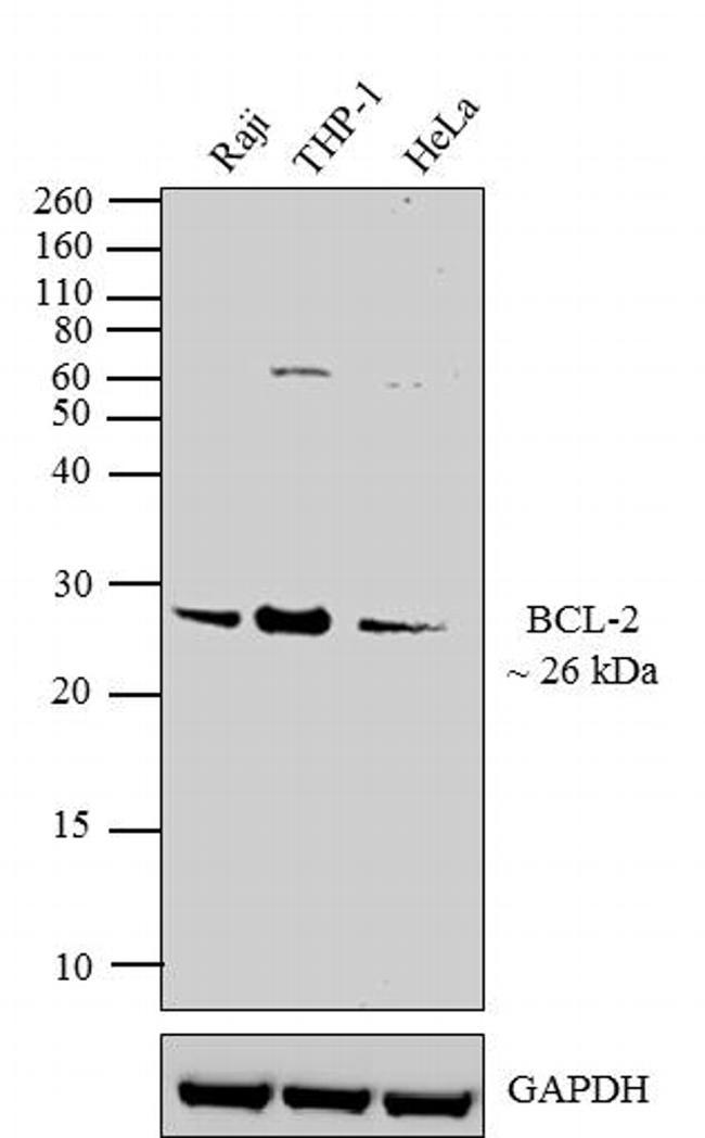
- Western blot analysis was performed on whole cell extracts (30 µg lysate) of Raji (Lane 1), THP-1 (Lane 2) and HeLa (Lane 3). The blots were probed with Anti-BCL-2 Mouse Monoclonal Antibody (Product # MA5-13666, 0.5-2 µg/mL) and detected by chemiluminescence using Goat anti-Mouse IgG (H+L) Secondary Antibody, HRP conjugate (Product # 62-6520, 1:4000 dilution). A 26 kDa band corresponding to BCL-2 was observed across cell lines tested. Known quantity of protein samples were electrophoresed using Novex® NuPAGE® 12 % Bis-Tris gel (Product # NP0342BOX), XCell SureLock™ Electrophoresis System (Product # EI0002) and Novex® Sharp Pre-Stained Protein Standard (Product # LC5800). Resolved proteins were then transferred onto a nitrocellulose membrane with iBlot® 2 Dry Blotting System (Product # IB21001). The membrane was probed with the relevant primary and secondary Antibody following blocking with 5 % skimmed milk. Chemiluminescent detection was performed using Novex® ECL Chemiluminescent Substrate Reagent Kit (Product # WP20005).
Supportive validation
- Submitted by
- Invitrogen Antibodies (provider)
- Main image
- Experimental details
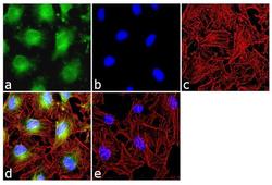
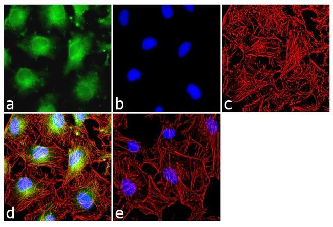
- Immunofluorescence analysis of BCL-2 was done on 70% confluent log phase HeLa cells. The cells were fixed with 4% paraformaldehyde for 10 minutes, permeabilized with 0.1% Triton™ X-100 for 10 minutes, and blocked with 1% BSA for 1 hour at room temperature. The cells were labeled BCL-2 (8C8) Mouse Monoclonal Antibody (MA513666) at 2ug/ml in 0.1% BSA and incubated for 3 hours at room temperature and then labeled with Goat anti-Mouse IgG (H+L) Superclonal™ Secondary Antibody, Alexa Fluor® 488 conjugate (Product # A28175) at a dilution of 1:2000 for 45 minutes at room temperature (Panel a: green). Nuclei (Panel b: blue) were stained with SlowFade® Gold Antifade Mountant with DAPI (S36938). F-actin (Panel c: red) was stained with Alexa Fluor® 555 Rhodamine Phalloidin (Product # R415, 1:300). Panel d is a merged image showing cytoplasmic and nuclear localization. Panel e is a no primary antibody control. The images were captured at 60X magnification.
Supportive validation
- Submitted by
- Invitrogen Antibodies (provider)
- Main image

- Experimental details
- Formalin-fixed, paraffin-embedded human tonsil stained with Bcl-2 alpha antibody using peroxidase-conjugate and DAB chromogen. Note absence of staining of reactive cells in the germinal center.
