Explore
 Validate
 Learn
Learn Western blot
Antibody data
- Antibody Data
- Antigen structure
- References [2]
- Comments [0]
- Validations
- Western blot [1]
- Immunocytochemistry [4]
- Flow cytometry [2]
- Other assay [1]
Submit
Validation data
Reference
Comment
Report error
- Product number
- MA5-15046 - Provider product page

- Provider
- Invitrogen Antibodies
- Product name
- Phospho-Bcl-2 (Ser70) Monoclonal Antibody (R.65.1)
- Antibody type
- Monoclonal
- Antigen
- Synthetic peptide
- Description
- It is not recommended to aliquot this antibody. This antibody is not cross-reactive with nonphosphorylated Bcl-2 at endogenous levels or with other Bcl-2 family members.
- Reactivity
- Human
- Host
- Rabbit
- Isotype
- IgG
- Antibody clone number
- R.65.1
- Vial size
- 100 μL
- Concentration
- 57 μg/mL
- Storage
- -20°C
Submitted references Anthocyanins from Hibiscus syriacus L. Inhibit Oxidative Stress-Mediated Apoptosis by Activating the Nrf2/HO-1 Signaling Pathway.
NF-κB RNAi decreases the Bax/Bcl-2 ratio and inhibits TNF-α-induced apoptosis in human alveolar epithelial cells.
Molagoda IMN, Lee KT, Choi YH, Kim GY
Antioxidants (Basel, Switzerland) 2020 Jan 3;9(1)
Antioxidants (Basel, Switzerland) 2020 Jan 3;9(1)
NF-κB RNAi decreases the Bax/Bcl-2 ratio and inhibits TNF-α-induced apoptosis in human alveolar epithelial cells.
Li L, Wu W, Huang W, Hu G, Yuan W, Li W
Inflammation research : official journal of the European Histamine Research Society ... [et al.] 2013 Apr;62(4):387-97
Inflammation research : official journal of the European Histamine Research Society ... [et al.] 2013 Apr;62(4):387-97
No comments: Submit comment
Supportive validation
- Submitted by
- Invitrogen Antibodies (provider)
- Main image

- Experimental details
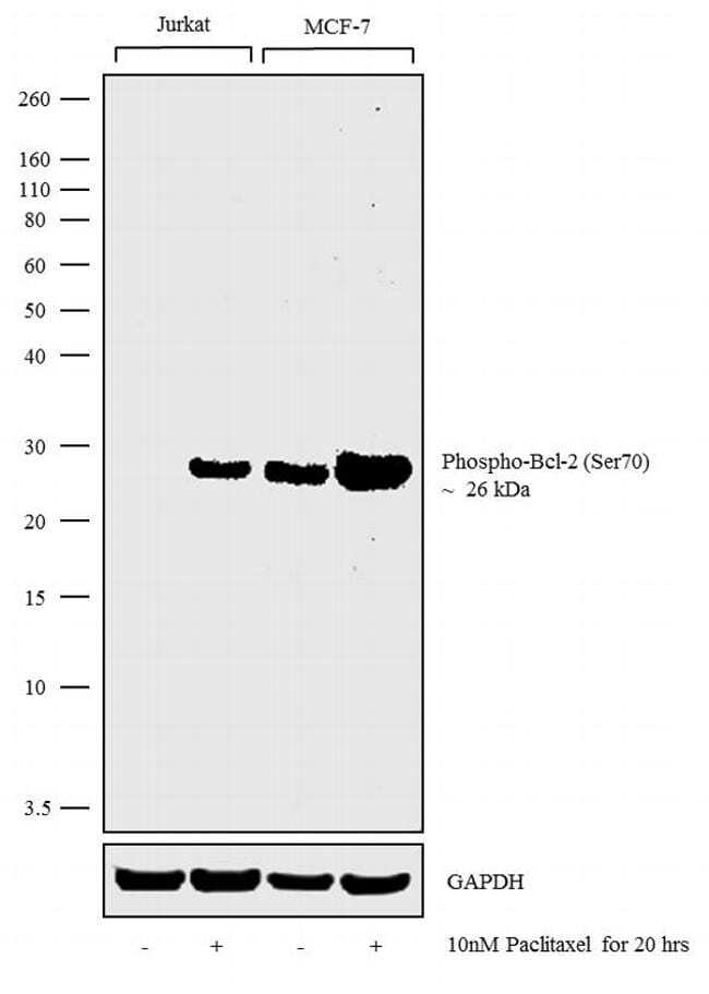
- Western blot analysis was performed on whole cell extracts (30 µg lysate) of Jurkat (Lane 1), Jurkat treated with Paclitaxel (10 nM for 20 hours) (Lane 2), MCF-7 (Lane 3) and MCF-7 treated with Paclitaxel (10 nM for 20 hours) (Lane 4). The blot was probed with Anti-Phospho-Bcl-2 (Ser70) Monoclonal Antibody (Product # MA5-15046, 1:1000 dilution) and detected by chemiluminescence using Goat anti-Rabbit IgG (Heavy Chain) Superclonal™ Secondary Antibody, HRP conjugate (Product # A27036, 0.25 µg/mL, 1:4000 dilution). A 26 kDa band corresponding to Phospho-Bcl-2 (Ser70) was observed to be increased in Paclitaxel treated cell lines tested.
Supportive validation
- Submitted by
- Invitrogen Antibodies (provider)
- Main image

- Experimental details
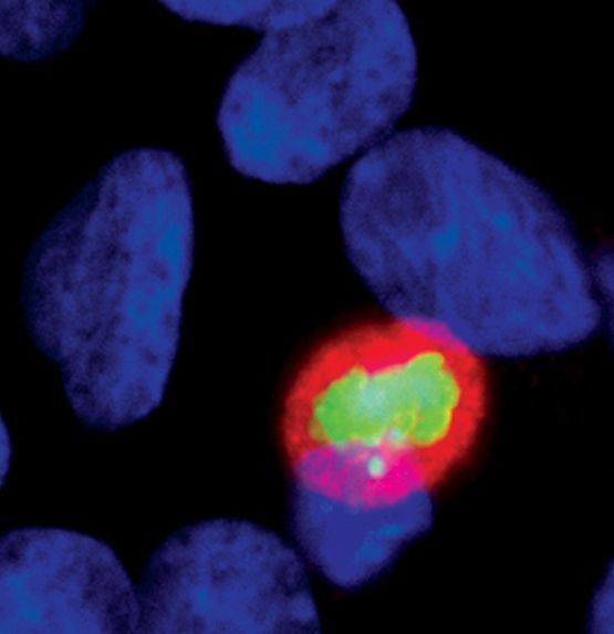
- Immunofluorescent analysis of Phospho-BCL-2 pSer70 in untreated SH-SY5Y cells using a Phospho-BCL-2 pSer70 monoclonal antibody (Product # MA5-15046) (green). Actin filaments are labeled with a fluorescent red phalloidin. DNA is labeled using a fluorescent blue dye.
- Submitted by
- Invitrogen Antibodies (provider)
- Main image

- Experimental details
- Immunofluorescent analysis of Phospho-Bcl-2 (Ser70) using a monoclonal antibody (Product # MA5-15046).

- Submitted by
- Invitrogen Antibodies (provider)
- Main image
- Experimental details
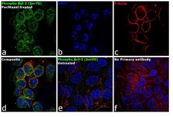
- Immunofluorescence analysis of Phospho-Bcl-2 (Ser70) was performed using 70% confluent log phase A-431 cells treated with 1mM of Paclitaxel for 20 hours. The cells were fixed with 4% paraformaldehyde for 10 minutes, permeabilized with 0.1% Triton™ X-100 for 15 minutes, and blocked with 1% BSA for 1 hour at room temperature. The cells were labeled with Phospho-Bcl-2 (Ser70) Rabbit Monoclonal Antibody (Product # PA5-15046) at 1:100 dilution in 0.1% BSA, incubated at 4 degree Celsius overnight and then labeled with Goat anti-Rabbit IgG (H+L) Superclonal™ Secondary Antibody, Alexa Fluor® 488 conjugate (Product # A27034) at a dilution of 1:2000 for 45 minutes at room temperature (Panel a: green). Nuclei (Panel b: blue) were stained with SlowFade® Gold Antifade Mountant with DAPI (Product # S36938). F-actin (Panel c: red) was stained with Rhodamine Phalloidin (Product # R415, 1:300). Panel d represents the merged image showing cytoplasmic localization. Panel e shows untreated cells with reduced signal. Panel f represents control cells with no primary antibody to assess background. The images were captured at 60X magnification.

- Submitted by
- Invitrogen Antibodies (provider)
- Main image

- Experimental details
- Immunofluorescence analysis of Phospho-Bcl-2 (Ser70) was performed using 70% confluent log phase A-431 cells treated with 1mM of Paclitaxel for 20 hours. The cells were fixed with 4% paraformaldehyde for 10 minutes, permeabilized with 0.1% Triton™ X-100 for 15 minutes, and blocked with 1% BSA for 1 hour at room temperature. The cells were labeled with Phospho-Bcl-2 (Ser70) Rabbit Monoclonal Antibody (Product # PA5-15046) at 1:100 dilution in 0.1% BSA, incubated at 4 degree Celsius overnight and then labeled with Goat anti-Rabbit IgG (Heavy Chain) Superclonal™ Secondary Antibody, Alexa Fluor® 488 conjugate (Product # A27034) at a dilution of 1:2000 for 45 minutes at room temperature (Panel a: green). Nuclei (Panel b: blue) were stained with SlowFade® Gold Antifade Mountant with DAPI (Product # S36938). F-actin (Panel c: red) was stained with Rhodamine Phalloidin (Product # R415, 1:300). Panel d represents the merged image showing cytoplasmic localization. Panel e shows untreated cells with reduced signal. Panel f represents control cells with no primary antibody to assess background. The images were captured at 60X magnification.

Supportive validation
- Submitted by
- Invitrogen Antibodies (provider)
- Main image
- Experimental details
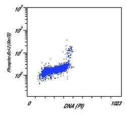
- Flow cytometric analysis of Phospho-BCL-2 pSer70 in Jurkat cells using a Phospho-BCL-2 pSer70 monoclonal antibody (Product # MA5-15046) versus propidium iodide (DNA content).

- Submitted by
- Invitrogen Antibodies (provider)
- Main image

- Experimental details
- Flow cytometric analysis of Phospho-BCL-2 pSer70 in Jurkat cells using a Phospho-BCL-2 pSer70 monoclonal antibody (Product # MA5-15046) versus propidium iodide (DNA content).

Supportive validation
- Submitted by
- Invitrogen Antibodies (provider)
- Main image

- Experimental details
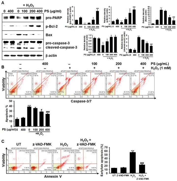
- Figure 3 PS protects HaCaT keratinocytes against H 2 O 2 -mediated apoptosis by modulating apoptosis-related proteins. HaCaT keratinocytes were seeded at a density of 1 x 10 4 cells/mL and pretreated with the indicated concentrations of PS (0-400 mug/mL) for 20 h prior to treatment with 1000 muM H 2 O 2 for 4 h. ( A ) The cells were lysed with radioimmuno precipitation assay lysis buffer and Western blotting was performed to identify poly (ADP-ribose) polymerase (PARP, 116 kDa), p-Bcl-2 (26 kDa), Bax (23 kDa), and caspase-3 (32 kDa). beta-Actin (43 kDa) was used as the internal control. Densitometry analysis is shown. ( B ) HaCaT keratinocytes were incubated in fluorogenic Muse (r) Caspase-3/7 reagent for 30 min at 37 degC and then incubated with a dead cell stain, 7-AAD, at 37 degC for 20 min. Caspase-3/7 + apoptotic cell populations were measured by using a Muse (r) Cell Analyzer. ( C ) In a parallel experiment, HaCaT keratinocytes were pretreated with 10 muM pan-caspase inhibitor, z-VAD-FMK, for 2 h prior to exposure to H 2 O 2 for 4 h. Early/late apoptotic populations were then measured by flow cytometry using Muse (r) Annexin V & Dead Cell Kit. Graphs represent caspase-3/7 + apoptotic cell populations ( B ) and proportion of cells in early/late apoptosis ( C ). All data are averaged from three independent experiments and presented as the mean +- the standard error of the median [*** p < 0.001 and ** p < 0.01 vs. the untreated group (UT) and ### p < 0.001 a