 Explore
 Validate
 Learn
Learn Western blot
 Immunocytochemistry
Antibody data
- Antibody Data
- Antigen structure
- References [8]
- Comments [0]
- Validations
- Immunocytochemistry [2]
- Immunohistochemistry [3]
- Other assay [8]
Submit
Validation data
Reference
Comment
Report error
- Product number
- PA5-20068 - Provider product page

- Provider
- Invitrogen Antibodies
- Product name
- Bcl-2 Polyclonal Antibody
- Antibody type
- Polyclonal
- Antigen
- Synthetic peptide
- Description
- A suggested positive control is Daudi cell lysate. PA5-20068 can be used with blocking peptide PEP-0187. The PA5-20068 immunogen is located within the first 50 amino acids of Bcl-2. Predicted molecular ~ 26kD. In Western blot applications, this antibody has been observed to detect a band at: 26 kD. Predicted species reactivity based on immunogen sequence: Bovine: (100%), Rat: (100%), Chicken: (93%)
- Reactivity
- Human, Mouse
- Host
- Rabbit
- Isotype
- IgG
- Vial size
- 100 μg
- Concentration
- 1 mg/mL
- Storage
- Maintain refrigerated at 2-8°C for up to 3 months. For long term storage store at -20°C
Submitted references Beneficial effect of trimetazidine on folic acid-induced acute kidney injury in mice: Role of HIF-1α/HO-1.
The protective effect of Ganoderma lucidum on testicular torsion/detorsion-induced ischemia-reperfusion (I/R) injury.
BDA-366, a putative Bcl-2 BH4 domain antagonist, induces apoptosis independently of Bcl-2 in a variety of cancer cell models.
Small-Dose Sunitinib Modulates p53, Bcl-2, STAT3, and ERK1/2 Pathways and Protects against Adenine-Induced Nephrotoxicity.
Erlotinib can halt adenine induced nephrotoxicity in mice through modulating ERK1/2, STAT3, p53 and apoptotic pathways.
The AKT/BCL-2 Axis Mediates Survival of Uterine Leiomyoma in a Novel 3D Spheroid Model.
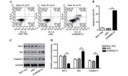
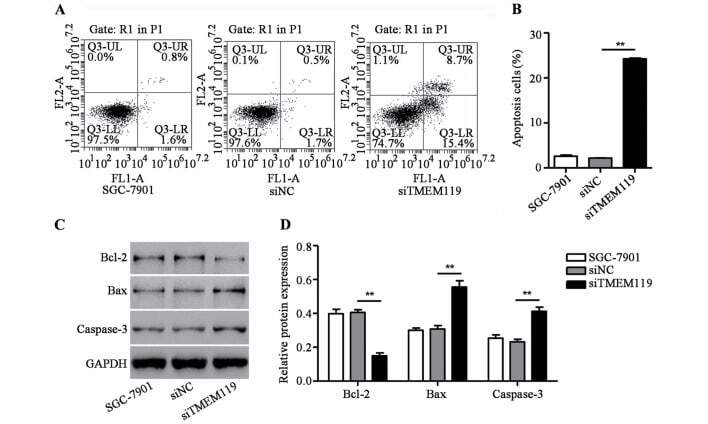
TMEM119 silencing inhibits cell viability and causes the apoptosis of gastric cancer SGC-7901 cells.
Inhibition of endoplasmic reticulum stress in high-fat-diet-induced obese C57BL/6 mice: Efficacy of a novel extract from mulberry (Morus alba) leaves fermented with Cordyceps militaris.
Abdelrahman RS, Abdelsalam RA, Zaghloul MS
Journal of biochemical and molecular toxicology 2022 May;36(5):e23011
Journal of biochemical and molecular toxicology 2022 May;36(5):e23011
The protective effect of Ganoderma lucidum on testicular torsion/detorsion-induced ischemia-reperfusion (I/R) injury.
Doğan G, İpek H
Acta cirurgica brasileira 2020;35(1):e202000103
Acta cirurgica brasileira 2020;35(1):e202000103
BDA-366, a putative Bcl-2 BH4 domain antagonist, induces apoptosis independently of Bcl-2 in a variety of cancer cell models.
Vervloessem T, Sasi BK, Xerxa E, Karamanou S, Kale J, La Rovere RM, Chakraborty S, Sneyers F, Vogler M, Economou A, Laurenti L, Andrews DW, Efremov DG, Bultynck G
Cell death & disease 2020 Sep 17;11(9):769
Cell death & disease 2020 Sep 17;11(9):769
Small-Dose Sunitinib Modulates p53, Bcl-2, STAT3, and ERK1/2 Pathways and Protects against Adenine-Induced Nephrotoxicity.
Saleh MA, Awad AM, Ibrahim TM, Abu-Elsaad NM
Pharmaceuticals (Basel, Switzerland) 2020 Nov 17;13(11)
Pharmaceuticals (Basel, Switzerland) 2020 Nov 17;13(11)
Erlotinib can halt adenine induced nephrotoxicity in mice through modulating ERK1/2, STAT3, p53 and apoptotic pathways.
Awad AM, Saleh MA, Abu-Elsaad NM, Ibrahim TM
Scientific reports 2020 Jul 13;10(1):11524
Scientific reports 2020 Jul 13;10(1):11524
The AKT/BCL-2 Axis Mediates Survival of Uterine Leiomyoma in a Novel 3D Spheroid Model.
Vidimar V, Chakravarti D, Bulun SE, Yin P, Nowak R, Wei JJ, Kim JJ
Endocrinology 2018 Mar 1;159(3):1453-1462
Endocrinology 2018 Mar 1;159(3):1453-1462
TMEM119 silencing inhibits cell viability and causes the apoptosis of gastric cancer SGC-7901 cells.
Zheng P, Wang W, Ji M, Zhu Q, Feng Y, Zhou F, He Q
Oncology letters 2018 Jun;15(6):8281-8286
Oncology letters 2018 Jun;15(6):8281-8286
Inhibition of endoplasmic reticulum stress in high-fat-diet-induced obese C57BL/6 mice: Efficacy of a novel extract from mulberry (Morus alba) leaves fermented with Cordyceps militaris.
Lee MR, Bae SJ, Kim JE, Song BR, Choi JY, Park JJ, Park JW, Kang MJ, Choi HJ, Choi YW, Kim KM, Hwang DY
Laboratory animal research 2018 Dec;34(4):288-294
Laboratory animal research 2018 Dec;34(4):288-294
No comments: Submit comment
Supportive validation
- Submitted by
- Invitrogen Antibodies (provider)
- Main image
- Experimental details
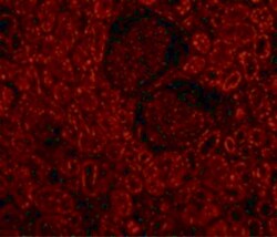
- Immunofluorescent analysis of 4% paraformaldehyde-fixed Human Kidney Cells labeling Bcl-2 with Bcl-2 Polyclonal Antibody (Product # PA5-20068) at 10 µg/mL, followed by goat anti-rabbit IgG secondary antibody at 1:500 dilution (red).
- Submitted by
- Invitrogen Antibodies (provider)
- Main image

- Experimental details
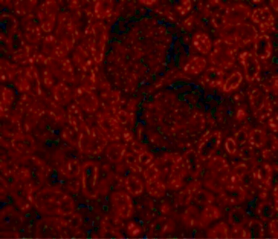
- Immunofluorescent analysis of 4% paraformaldehyde-fixed Human Kidney Cells labeling Bcl-2 with Bcl-2 Polyclonal Antibody (Product # PA5-20068) at 10 µg/mL, followed by goat anti-rabbit IgG secondary antibody at 1:500 dilution (red).

Supportive validation
- Submitted by
- Invitrogen Antibodies (provider)
- Main image
- Experimental details
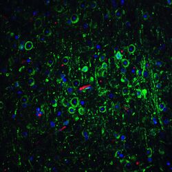
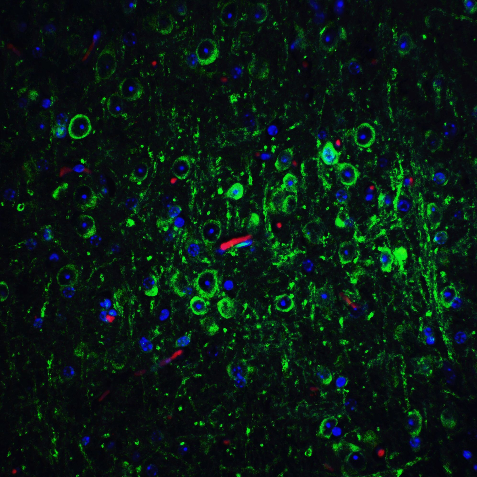
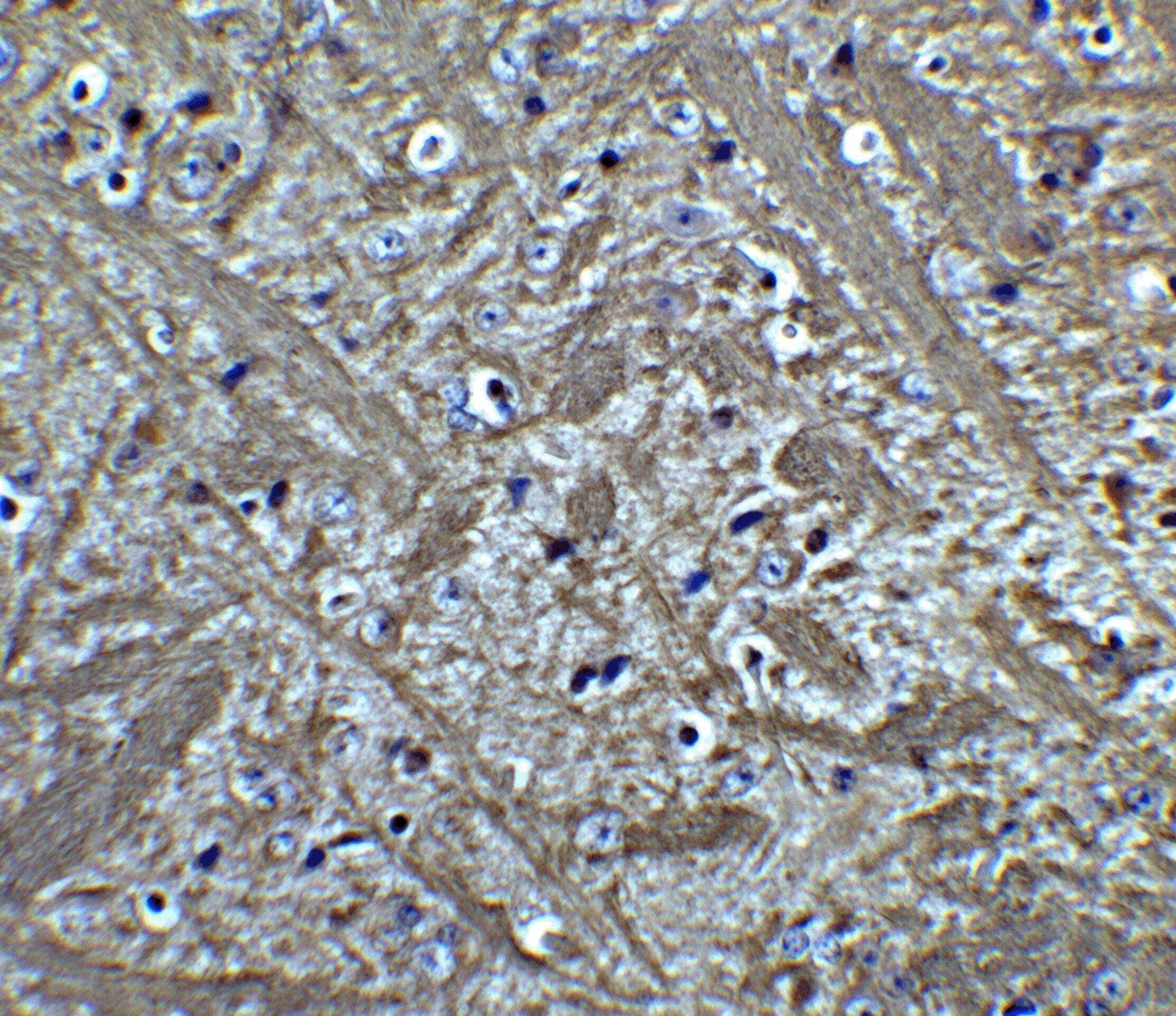
- Immunofluorescent analysis of 4% paraformaldehyde-fixed Mouse Brain Tissue labeling Bcl-2 with Bcl-2 Polyclonal Antibody (Product # PA5-20068) at 20 µg/mL, followed by goat anti-rabbit IgG secondary antibody at 1:500 dilution (green).
- Submitted by
- Invitrogen Antibodies (provider)
- Main image

- Experimental details
- Immunohistochemical analysis of paraffin-embedded Mouse Brain Tissue using Bcl-2 Polyclonal Antibody (Product # PA5-20068) at 5 µg/mL. Tissue was fixed with formaldehyde and blocked with 0.1 serum for 1 h at RT; antigen retrieval was by heat mediation with a citrate buffer (pH6). Samples were incubated with primary antibody overnight at 4˚C. A goat anti-rabbit IgG H&L (HRP) at 1/250 was used as secondary. Counter stained with Hematoxylin.
- Submitted by
- Invitrogen Antibodies (provider)
- Main image

- Experimental details
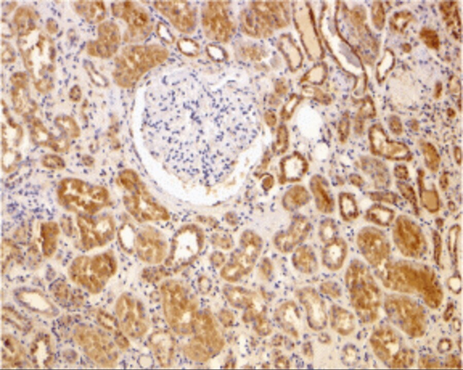
- Immunohistochemical analysis of paraffin-embedded Human Kidney using Bcl-2 Polyclonal Antibody (Product # PA5-20068) at 2 µg/mL. Tissue was fixed with formaldehyde and blocked with 0.1 serum for 1 h at RT; antigen retrieval was by heat mediation with a citrate buffer (pH6). Samples were incubated with primary antibody overnight at 4˚C. A goat anti-rabbit IgG H&L (HRP) at 1/250 was used as secondary. Counter stained with Hematoxylin.
Supportive validation
- Submitted by
- Invitrogen Antibodies (provider)
- Main image
- Experimental details
- Figure 3. TMEM119 silencing induced SGC-7901 cell apoptosis. After transfection of SGC-7901 cells with TMEM119-siRNA (siTMEM119), cell apoptosis was measured by (A and B) flow cytometry assay and the expression of Bcl-2, Bax as well as caspase-3 was measured by (C and D) western blot analysis. **P
- Submitted by
- Invitrogen Antibodies (provider)
- Main image
- Experimental details
- Figure 3 A: Sham group: The spermatogenic cells of the seminiferous tubules in the control group with strong mitotic activity, Sertoli cells with regular broad luminal faces. Normal membrane thickness, interstitial area, blood vessels and Leydig cells, PAS staining Bar 50 µm. B: T/D group: An increased thickness of basal membrane in tubules, pyknosis and deterioration in the nucleus of spermatogenic cells, lack of maturation in sperm cells and degeneration of Sertoli cells, hemorrhage and dilatation of blood vessels, an increase in connective tissue, degeneration of Leydig cells, PAS staining Bar 50 µm. C: T/D+G. lucidum : Decreased basal membrane thickness of tubules, degeneration and deterioration of spermatic cells in some tubules and mitotic increase of spermatic cells in many tubules, small hemorrhage in interstitial blood vessels, heterochromatin appearance of Leydig cells, PAS staining Bar 50 µm. D: Sham group: Negative VEGF expression in spermatogenetic and Sertoli cells in tubules, interstitial vascular endothelial cells and Leydig cells, VEGF staining Bar 50 µm. E: T/D group: Positive expression of VEGF on apical surfaces of spermatogenic cells and Sertoli cells in tubules, VEGF expression positive in endothelial cells and some inflammatory cells in interstitial blood vessels and also in Leydig cells, VEGF staining Bar 50 µm. F: T/D + G. lucidum group: positive VEGF expression in a few spermatogenic cells in some tubules, especially in
- Submitted by
- Invitrogen Antibodies (provider)
- Main image
- Experimental details
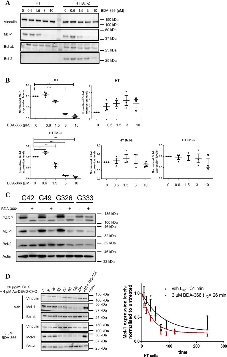
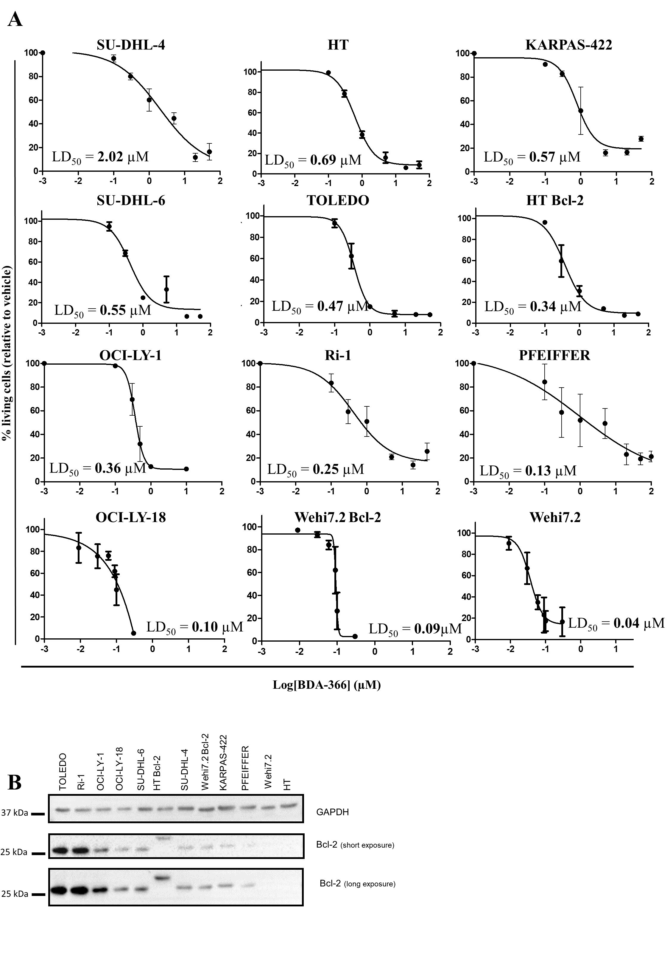
- Fig. 6 BDA-366 augments Mcl-1 turnover in a Bcl-2-independent manner. a Representative western blot of different Bcl-2 family members after BDA-366 treatment for 6 h in HT and HT overexpressing Bcl-2 positive (HT Bcl-2) DLBCL cells. Vinculin was used as a loading control. b Quantitative analysis of the different Bcl-2 family members (Mcl-1, Bcl-2, and Bcl-xL). Data are presented as average +- SD of three independent experiment ( N = 3) with * p < 0.05, ** p < 0.01, *** p < 0.001 obtained via a two-tailed paired t test. c Immunoblotting analysis of PARP cleavage (indicator for apoptosis), Mcl-1 and Bcl-2 levels after 48 h treatment of CLL cells with 2 uM BDA-366. Actin was used as loading control. d HT cells were incubated for different time points with 3 uM BDA-366 in combination with 20 µg/mL CHX and the capsase-3 inhibitor (Ac-DEVD-CHO, 4 uM) and subjected to Mcl-1 immunoblotting. The 4-hours time point was analyzed with or without proteasome inhibitor (MG-132, 20 uM). The protein concentrations relative to time 0 were fitted using a one-phase exponential-decay to calculate the half life ( t 1/2 ). Significance was measured via a paired t-test comparing vehicle versus BDA-366 treatment at each timepoint.
- Submitted by
- Invitrogen Antibodies (provider)
- Main image
- Experimental details
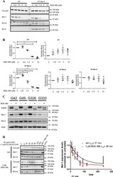
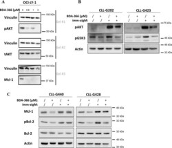
- Fig. 7 BDA-366 decreases the phosphorylation of AKT and Bcl-2. a Representative western blots of pAKT, tAKT, and Mcl-1 in OCI-LY-1 cells (LD 50 for BDA-366: 0.32 muM) treated for 6 h with 0-0.6-1-3 uM BDA-366. Vinculin was used as a loading control. One representative experiment out of 3 performed is shown. b BDA-366 inhibits BCR-induced AKT and GSK3 phosphorylation in primary CLL cells. Cells were pretreated with BDA-366 (2 muM) for 90 min prior to being stimulated with immobilized anti-IgM (imm-aIgM) for 30 min (2 x 10 7 beads coated with 20 mug goat anti-human IgM antibody per 1 x 10 7 cells) . c BDA-366 inhibits BCR-induced Mcl-1 upregulation and Bcl-2 phosphorylation in primary CLL cells. Cells were pretreated with BDA-366 (2 muM) as above and then stimulated for 24 h with imm-aIgM prior to harvesting for immunoblotting analysis.

- Submitted by
- Invitrogen Antibodies (provider)
- Main image

- Experimental details
- Supplemental Figure 2
- Submitted by
- Invitrogen Antibodies (provider)
- Main image

- Experimental details
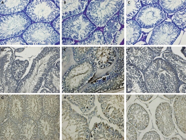
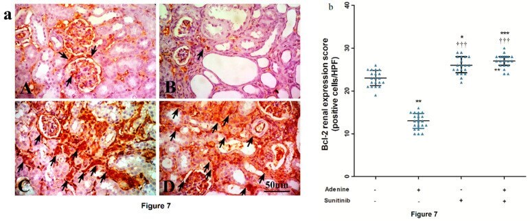
- Figure 7 ( a ) Representative immuno-stained kidney sections photographs showing an expression of B-cell lymphoma (Bcl)-2 (arrows) isolated from the ( A ) normal group, ( B ) adenine group, ( C ) sunitinib group, and ( D ) adenine+sunitinib group. ( b ) Effect of sunitinib on kidney expression scores of Bcl-2 in an adenine model of nephrotoxicity ( n = 8 mice/group). Significance: *, **, *** p < 0.05, 0.01, and 0.001 compared to the normal group; +++ p < 0.01, 0.001 compared to the adenine group.
- Submitted by
- Invitrogen Antibodies (provider)
- Main image

- Experimental details
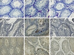
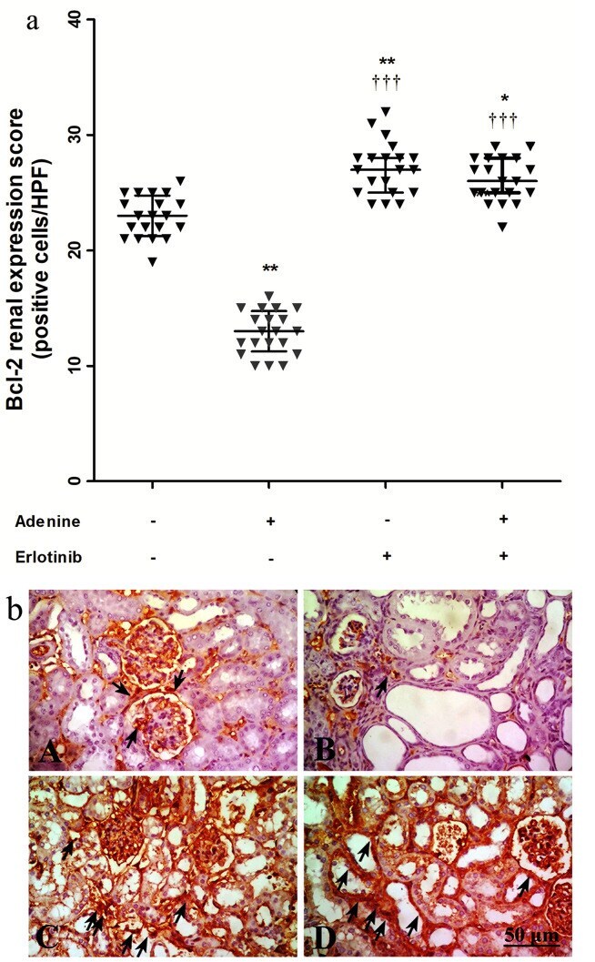
- Figure 5 ( a ) Effect of erlotinib (80 mg/kg) administration on kidney expression scores of B-cell lymphoma (Bcl)-2 in adenine induced nephrotoxicity in mice. Significance: *, ** P < 0.05, 0.01 compared to control group; ++++++ P < 0.001 compared to adenine group. ( b ) Representative photographs for immuno-stained kidney sections isolated from A: control group B: adenine group C: erlotinib group D: adenine + erlotinib group showing expression of Bcl-2 (arrows).
- Submitted by
- Invitrogen Antibodies (provider)
- Main image
- Experimental details
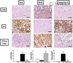
- 3 Figure Effect of trimetazidine (20 mg/kg) on kidney apoptotic markers induced by folic acid in mice. Upper panel: Representative photomicrographs of immunohistochemical detection of proapoptotic Bax (left panel); antiapoptotic Bcl2 (middle panel); caspase-3 (right panel) (x100, bar = 100 um). Lower panel: bar graphs showing area of proapoptotic Bax (left panel); antiapoptotic Bcl2 (middle panel); caspase-3 (right panel) positive staining statistical significances of data (mean +- SEM, n = 4) were denoted as *, $ ( p < 0.05) from control groups and folic acid groups, respectively, using one-way ANOVA followed by Tukey-Kramer multiple comparisons test. ANOVA, analysis of variance; CON, control; FA, folic acid TMZ, trimetazidine
