 Explore
 Validate
 Learn
Learn Western blot
 Immunocytochemistry
Antibody data
- Antibody Data
- Antigen structure
- References [13]
- Comments [0]
- Validations
- Immunocytochemistry [2]
- Immunohistochemistry [4]
- Other assay [8]
Submit
Validation data
Reference
Comment
Report error
- Product number
- PA5-27094 - Provider product page

- Provider
- Invitrogen Antibodies
- Product name
- Bcl-2 Polyclonal Antibody
- Antibody type
- Polyclonal
- Antigen
- Synthetic peptide
- Description
- Recommended positive controls: THP-1, HL-60, mouse spleen, PA-1, OKT3. Predicted reactivity: Mouse (100%), Rat (100%), Dog (100%), Cat (100%). Store product as a concentrated solution. Centrifuge briefly prior to opening the vial.
- Reactivity
- Human, Mouse, Rat
- Host
- Rabbit
- Isotype
- IgG
- Vial size
- 100 μL
- Concentration
- 0.2 mg/mL
- Storage
- Store at 4°C short term. For long term storage, store at -20°C, avoiding freeze/thaw cycles.
Submitted references Preventive Effect of Limosilactobacillus fermentum SCHY34 on Lead Acetate-Induced Neurological Damage in SD Rats.
Infarct-preconditioning exosomes of umbilical cord mesenchymal stem cells promoted vascular remodeling and neurological recovery after stroke in rats.
Orientin, a Bio-Flavonoid from Trigonella hamosa L., Regulates COX-2/PGE-2 in A549 Cell Lines via miR-26b and miR-146a.
Astragaloside IV protects against ischemia/reperfusion (I/R)-induced kidney injury based on the Keap1-Nrf2/ARE signaling pathway.
A Nano-Liposomal Formulation of Caffeic Acid Phenethyl Ester Modulates Nrf2 and NF-κβ Signaling and Alleviates Experimentally Induced Acute Pancreatitis in a Rat Model.
Cardiac Protective Effect of Kirenol against Doxorubicin-Induced Cardiac Hypertrophy in H9c2 Cells through Nrf2 Signaling via PI3K/AKT Pathways.
Date Palm Pollen Extract Avert Doxorubicin-Induced Cardiomyopathy Fibrosis and Associated Oxidative/Nitrosative Stress, Inflammatory Cascade, and Apoptosis-Targeting Bax/Bcl-2 and Caspase-3 Signaling Pathways.
Human urine-derived stem cells protect against renal ischemia/reperfusion injury in a rat model via exosomal miR-146a-5p which targets IRAK1.
An iPSC-Derived Neuron Model of CLN3 Disease Facilitates Small Molecule Phenotypic Screening.
Human umbilical cord-derived mesenchymal stem cells and human cord blood mononuclear cells protect against cisplatin-induced acute kidney injury in rat models.
Kaempferol Inhibits Zearalenone-Induced Oxidative Stress and Apoptosis via the PI3K/Akt-Mediated Nrf2 Signaling Pathway: In Vitro and In Vivo Studies.
Molecular mechanisms underlying histological and biochemical changes induced by nitrate in rat liver and the efficacy of S-Allylcysteine.
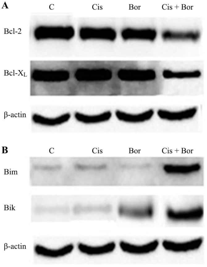
Synergistic effects of cisplatin and proteasome inhibitor bortezomib on human bladder cancer cells.
Long X, Wu H, Zhou Y, Wan Y, Kan X, Gong J, Zhao X
Frontiers in nutrition 2022;9:852012
Frontiers in nutrition 2022;9:852012
Infarct-preconditioning exosomes of umbilical cord mesenchymal stem cells promoted vascular remodeling and neurological recovery after stroke in rats.
Ye YC, Chang ZH, Wang P, Wang YW, Liang J, Chen C, Wang JJ, Sun HT, Wang Y, Li XH
Stem cell research & therapy 2022 Jul 28;13(1):378
Stem cell research & therapy 2022 Jul 28;13(1):378
Orientin, a Bio-Flavonoid from Trigonella hamosa L., Regulates COX-2/PGE-2 in A549 Cell Lines via miR-26b and miR-146a.
Khalil HE, Ibrahim HM, Ahmed EA, Emeka PM, Alhaider IA
Pharmaceuticals (Basel, Switzerland) 2022 Jan 27;15(2)
Pharmaceuticals (Basel, Switzerland) 2022 Jan 27;15(2)
Astragaloside IV protects against ischemia/reperfusion (I/R)-induced kidney injury based on the Keap1-Nrf2/ARE signaling pathway.
Su Y, Xu J, Chen S, Feng J, Li J, Lei Z, Qiao L, Wang Y, Zeng D
Translational andrology and urology 2022 Aug;11(8):1177-1188
Translational andrology and urology 2022 Aug;11(8):1177-1188
A Nano-Liposomal Formulation of Caffeic Acid Phenethyl Ester Modulates Nrf2 and NF-κβ Signaling and Alleviates Experimentally Induced Acute Pancreatitis in a Rat Model.
Shahin NN, Shamma RN, Ahmed IS
Antioxidants (Basel, Switzerland) 2022 Aug 7;11(8)
Antioxidants (Basel, Switzerland) 2022 Aug 7;11(8)
Cardiac Protective Effect of Kirenol against Doxorubicin-Induced Cardiac Hypertrophy in H9c2 Cells through Nrf2 Signaling via PI3K/AKT Pathways.
Alzahrani AM, Rajendran P, Veeraraghavan VP, Hanieh H
International journal of molecular sciences 2021 Mar 23;22(6)
International journal of molecular sciences 2021 Mar 23;22(6)
Date Palm Pollen Extract Avert Doxorubicin-Induced Cardiomyopathy Fibrosis and Associated Oxidative/Nitrosative Stress, Inflammatory Cascade, and Apoptosis-Targeting Bax/Bcl-2 and Caspase-3 Signaling Pathways.
Elblehi SS, El-Sayed YS, Soliman MM, Shukry M
Animals : an open access journal from MDPI 2021 Mar 20;11(3)
Animals : an open access journal from MDPI 2021 Mar 20;11(3)
Human urine-derived stem cells protect against renal ischemia/reperfusion injury in a rat model via exosomal miR-146a-5p which targets IRAK1.
Li X, Liao J, Su X, Li W, Bi Z, Wang J, Su Q, Huang H, Wei Y, Gao Y, Li J, Liu L, Wang C
Theranostics 2020;10(21):9561-9578
Theranostics 2020;10(21):9561-9578
An iPSC-Derived Neuron Model of CLN3 Disease Facilitates Small Molecule Phenotypic Screening.
Kinarivala N, Morsy A, Patel R, Carmona AV, Sajib MS, Raut S, Mikelis CM, Al-Ahmad A, Trippier PC
ACS pharmacology & translational science 2020 Oct 9;3(5):931-947
ACS pharmacology & translational science 2020 Oct 9;3(5):931-947
Human umbilical cord-derived mesenchymal stem cells and human cord blood mononuclear cells protect against cisplatin-induced acute kidney injury in rat models.
Xu Q, Yan P, Duan XJ, Wu X, Chen XJ, Luo M, Peng JC, Feng LX, Liu J, Zhong HL, Cheng W, Zou QY, Duan SB
Experimental and therapeutic medicine 2020 Dec;20(6):145
Experimental and therapeutic medicine 2020 Dec;20(6):145
Kaempferol Inhibits Zearalenone-Induced Oxidative Stress and Apoptosis via the PI3K/Akt-Mediated Nrf2 Signaling Pathway: In Vitro and In Vivo Studies.
Rajendran P, Ammar RB, Al-Saeedi FJ, Mohamed ME, ElNaggar MA, Al-Ramadan SY, Bekhet GM, Soliman AM
International journal of molecular sciences 2020 Dec 28;22(1)
International journal of molecular sciences 2020 Dec 28;22(1)
Molecular mechanisms underlying histological and biochemical changes induced by nitrate in rat liver and the efficacy of S-Allylcysteine.
Kattaia AA, Abd El-Baset SA, Mohamed EM, Abdul-Maksou RS, Elfakharany YM
Ultrastructural pathology 2017 Jan-Feb;41(1):10-22
Ultrastructural pathology 2017 Jan-Feb;41(1):10-22
Synergistic effects of cisplatin and proteasome inhibitor bortezomib on human bladder cancer cells.
Konac E, Varol N, Kiliccioglu I, Bilen CY
Oncology letters 2015 Jul;10(1):560-564
Oncology letters 2015 Jul;10(1):560-564
No comments: Submit comment
Supportive validation
- Submitted by
- Invitrogen Antibodies (provider)
- Main image
- Experimental details
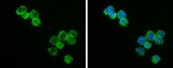
- Immunocytochemistry-Immunofluorescence analysis of Bcl-2 was performed in THP-1 cells fixed in 4% paraformaldehyde at RT for 15 min. Green: Bcl-2 Polyclonal Antibody (Product # PA5-27094) diluted at 1:500. Blue: Fluoroshield with DAPI.
- Submitted by
- Invitrogen Antibodies (provider)
- Main image

- Experimental details
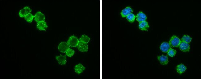
- Immunocytochemistry-Immunofluorescence analysis of Bcl-2 was performed in THP-1 cells fixed in 4% paraformaldehyde at RT for 15 min. Green: Bcl-2 Polyclonal Antibody (Product # PA5-27094) diluted at 1:500. Blue: Fluoroshield with DAPI.

Supportive validation
- Submitted by
- Invitrogen Antibodies (provider)
- Main image
- Experimental details
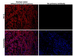
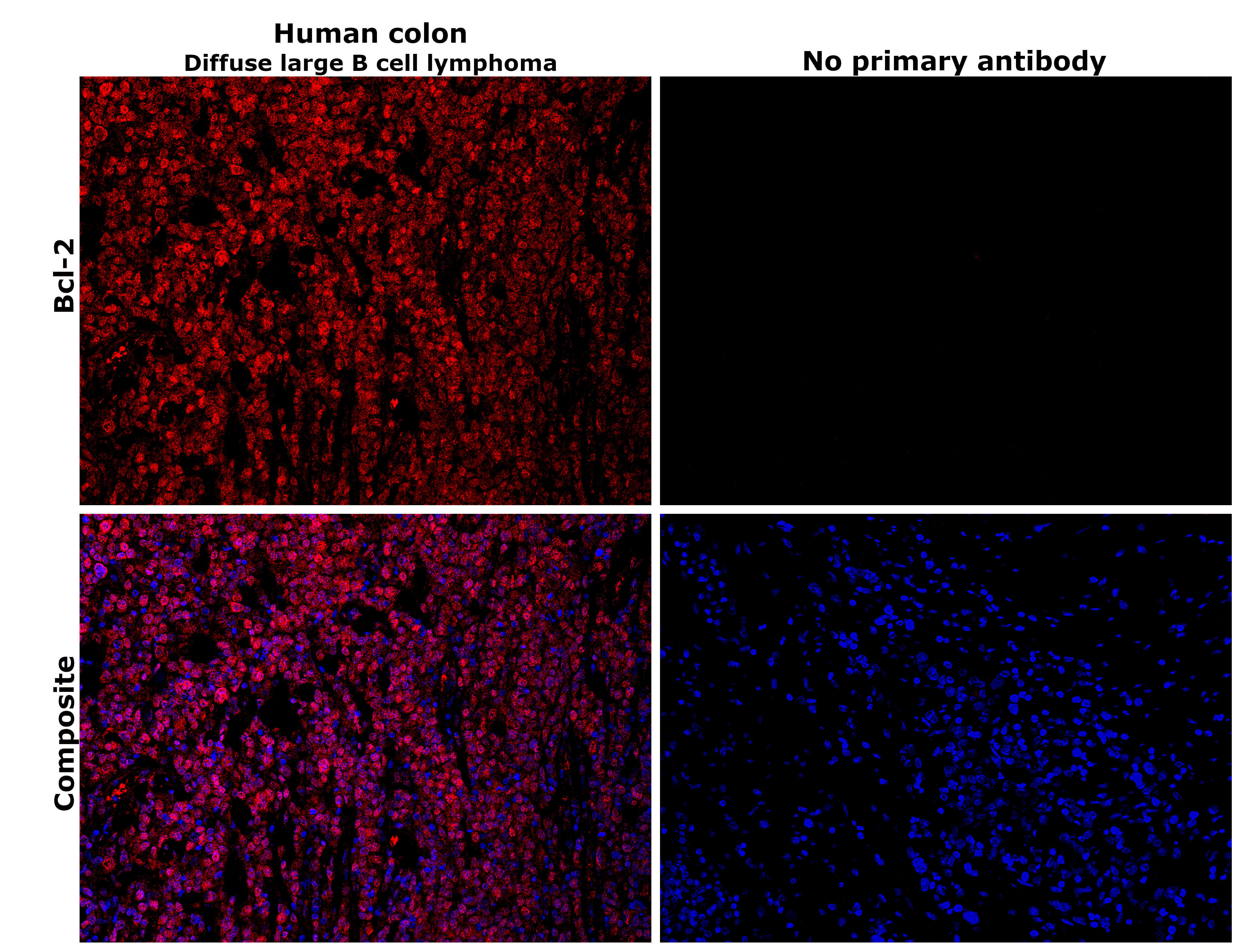
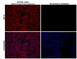
- Immunohistochemical analysis of Bcl-2 was performed using formalin-fixed paraffin-embedded human colon (Diffuse large B cell lymphoma) tissue sections. To expose the target protein, heat-induced epitope retrieval was performed on de-paraffinized sections using eBioscience™ IHC Antigen Retrieval Solution - Low pH (10X) (Product # 00-4955-58) diluted to 1X solution in water in a decloaking chamber at 110 degree Celsius for 15 minutes. Following antigen retrieval, the sections were blocked with 2% normal goat serum in 1X PBS for 45 minutes at room temperature and then probed with or without Bcl-2 Polyclonal Antibody (Product # PA5-27094) at 1:100 dilution in 0.1% normal goat serum overnight at 4 degree Celsius in a humidified chamber. Detection was performed using Goat anti-Rabbit IgG (H+L) Highly Cross-Adsorbed Secondary Antibody, Alexa Fluor™ Plus 647 (Product # A32733) at a dilution of 1:2,000 in 0.1% normal goat serum for 45 minutes at room temperature. ReadyProbes™ Tissue Autofluorescence Quenching Kit (Product # R37630) was used to quench autofluorescence from the tissues. Nuclei were stained with DAPI (Product # D1306) and the sections were mounted using ProLong™ Glass Antifade Mountant (Product # P36984). The images were captured on EVOS™ M7000 Imaging System (Product # AMF7000) at 20X magnification and externally deconvoluted.
- Submitted by
- Invitrogen Antibodies (provider)
- Main image
- Experimental details
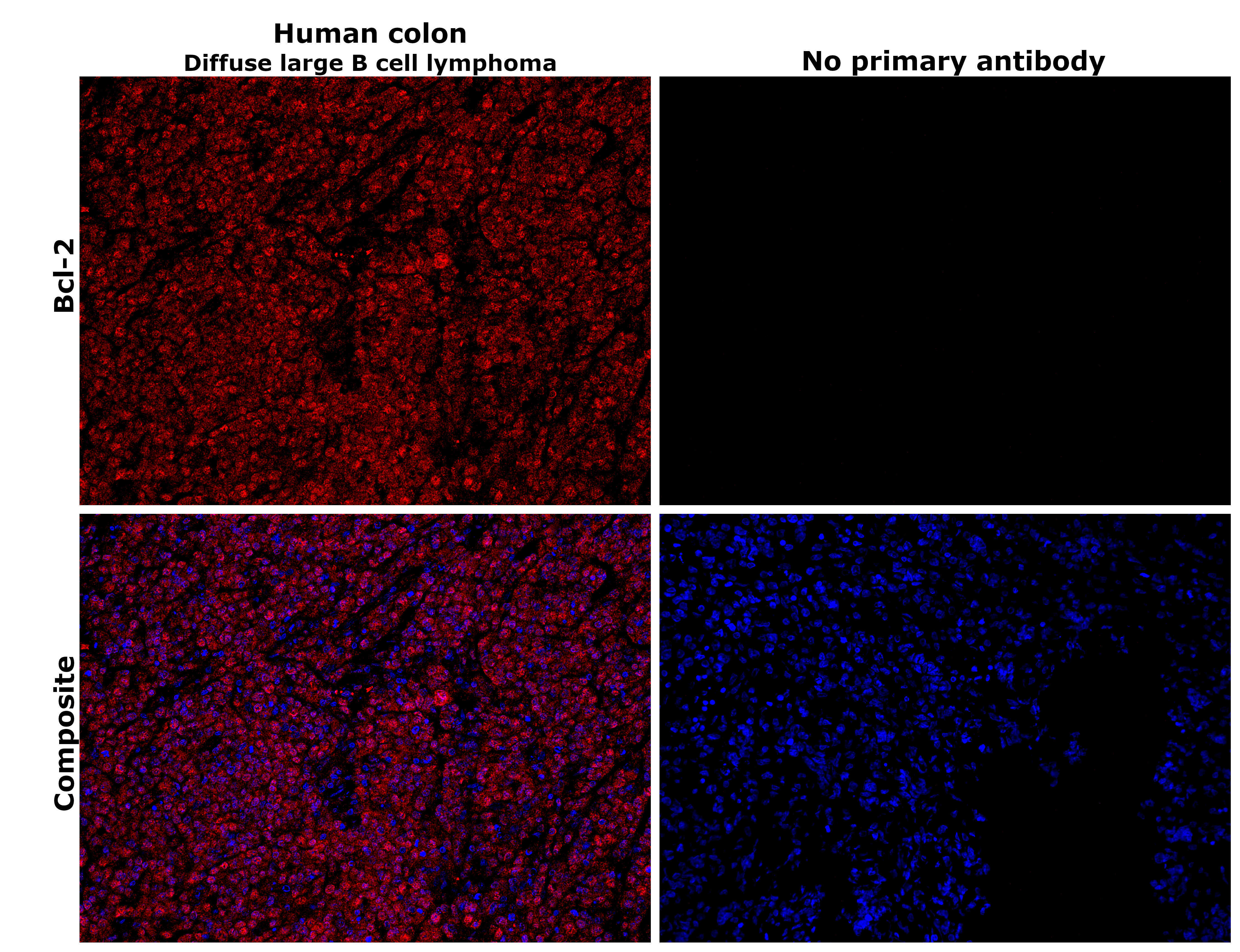
- Immunohistochemical analysis of Bcl-2 was performed using formalin-fixed paraffin-embedded human colon (Diffuse large B cell lymphoma) tissue sections. To expose the target protein, heat-induced epitope retrieval was performed on de-paraffinized sections using eBioscience™ IHC Antigen Retrieval Solution - High pH (10X) (Product # 00-4956-58) diluted to 1X solution in water in a decloaking chamber at 110 degree Celsius for 15 minutes. Following antigen retrieval, the sections were blocked with 2% normal goat serum in 1X PBS for 45 minutes at room temperature and then probed with or without Bcl-2 Polyclonal Antibody (Product # PA5-27094) at 1:100 dilution in 0.1% normal goat serum overnight at 4 degree Celsius in a humidified chamber. Detection was performed using Goat anti-Rabbit IgG (H+L) Highly Cross-Adsorbed Secondary Antibody, Alexa Fluor™ Plus 647 (Product # A32733) at a dilution of 1:2,000 in 0.1% normal goat serum for 45 minutes at room temperature. ReadyProbes™ Tissue Autofluorescence Quenching Kit (Product # R37630) was used to quench autofluorescence from the tissues. Nuclei were stained with DAPI (Product # D1306) and the sections were mounted using ProLong™ Glass Antifade Mountant (Product # P36984). The images were captured on EVOS™ M7000 Imaging System (Product # AMF7000) at 20X magnification and externally deconvoluted.
- Submitted by
- Invitrogen Antibodies (provider)
- Main image
- Experimental details
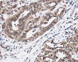
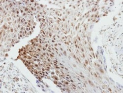
- Immunohistochemistry (Paraffin) analysis of Bcl-2 was performed in paraffin-embedded human cervix tissue using Bcl-2 Polyclonal Antibody (Product # PA5-27094) at a dilution of 1:500.
- Submitted by
- Invitrogen Antibodies (provider)
- Main image
- Experimental details
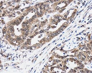
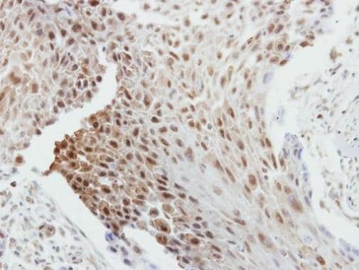
- Immunohistochemical analysis of paraffin-embedded human breast cancer, using Bcl-2 (Product # PA5-27094) antibody at 1:250 dilution. Antigen Retrieval: EDTA based buffer, pH 8.0, 15 min.
Supportive validation
- Submitted by
- Invitrogen Antibodies (provider)
- Main image

- Experimental details
- NULL
- Submitted by
- Invitrogen Antibodies (provider)
- Main image
- Experimental details
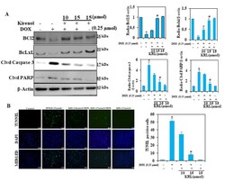
- Figure 6 KRL inhibits DOX-induced apoptosis in cardiomyocytes. ( A ) Cells were pretreated with 15 umol of KRL for 2 h followed by DOX (0.25 umol for 24 h). The expression levels of anti-apoptotic Bcl2 and Bcl-xL and apoptotic proteins cleaved caspase-3 activation and PARP cleavage were measured by the Western blot method. ( B ) Apoptotic nuclei were detected by terminal deoxynucleotidyl transferase dUTP nick-end labeling (TUNEL) staining and the nuclei were detected by DAPI staining, showing modulations in apoptotic levels with DOX against and with KRL. Scale bar indicated 100 um at 20x magnification. Data are represented as the mean +- SD of triplicate values ( n = 3) and * p < 0.05 represents significant variations compared with the control. # p < 0.05 represents significant variations as compared to DOX alone and KRL with DOX treatment groups.

- Submitted by
- Invitrogen Antibodies (provider)
- Main image
- Experimental details
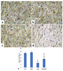
- Figure 7 Immunohistochemical staining of B-cell lymphoma-2 (Bcl-2) in cardiac cells of the experimental rats (IHC, x400). A control ( a ), DPPE-treated ( b ) DOX-treated ( c ) and DPP + DOX-treated ( d ) rats. ( e ) Quantification of Bcl-2 expression, the immunohistochemical staining of Bcl-2 was measured as area percent (%) across 10 different fields/section, n = 7 rat/group. Mean values were statistically different from the CTR ( # p < 0.05), DPPE (* p < 0.05), and DOX ( $ p < 0.05) groups.

- Submitted by
- Invitrogen Antibodies (provider)
- Main image

- Experimental details
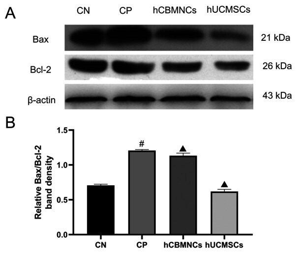
- Figure 5 Changes in Bax and Bcl-2 protein expression in the renal tissues of each group. (A) Protein levels of Bax and Bcl-2 were measured using western blotting. (B) Protein bands were quantified using Tanon 5200 Multi Image Analysis software and relative Bax/Bcl-2 band densities were measured. Data are presented as the mean +- SD (n=6/group). # P
- Submitted by
- Invitrogen Antibodies (provider)
- Main image
- Experimental details
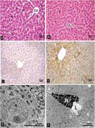
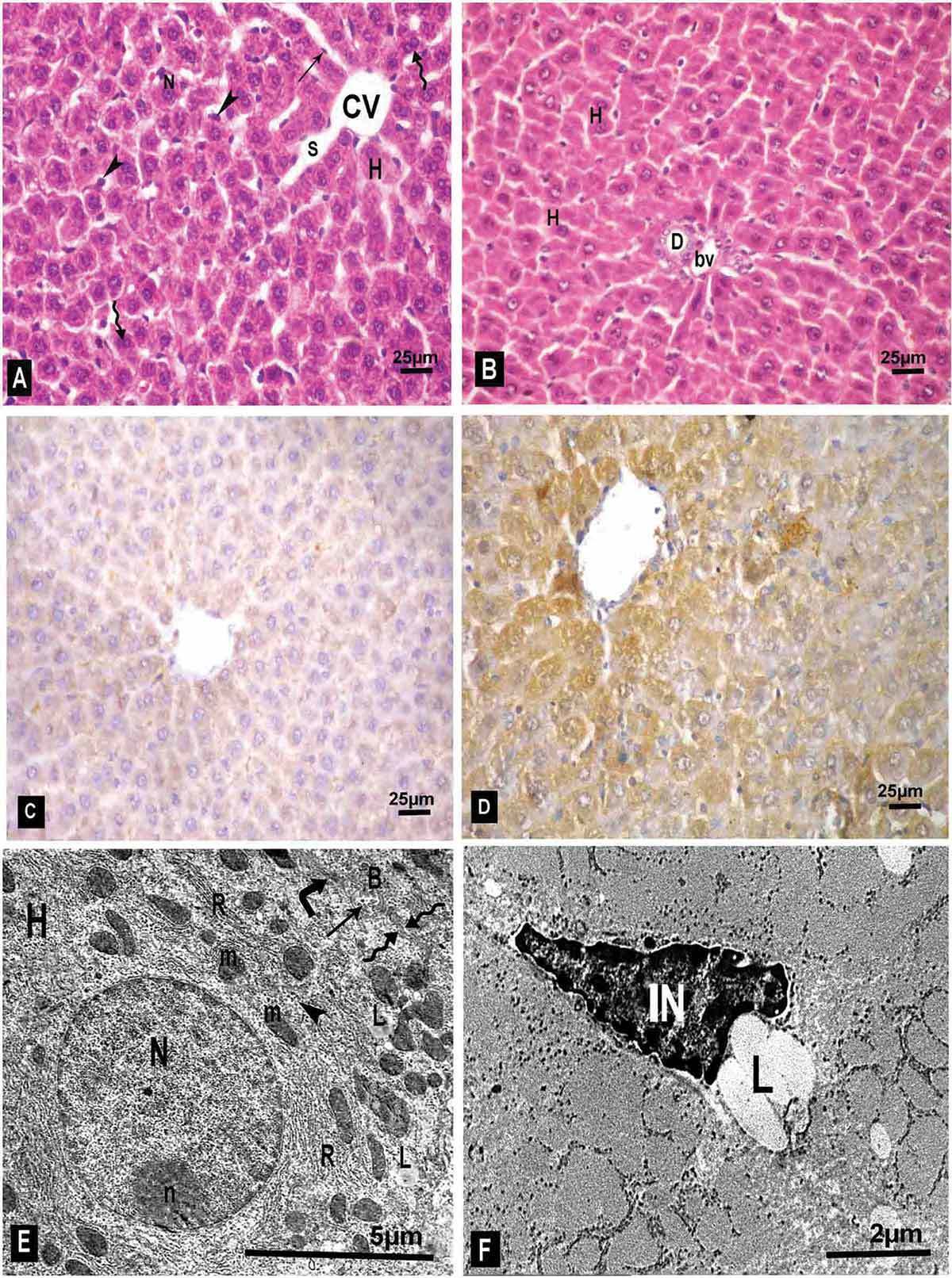
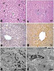
- Figure 1. A photomicrograph of sections in adult rat liver of the control group. (A & B): H&E-stained sections showing normal liver architecture with hepatocytes (H) radiating from central vein (CV), rounded vesicular nuclei (N), binucleated hepatocytes (wavy arrow), blood sinusoids (S) lined by endothelium (arrow) with some Kupffer cells (arrow head), Portal area with bile duct (D), blood vessel (bv). (C): immune stained section for heat shock protein (HSP) showing cytoplasmic positive immunoreactions in some hepatocytes. (D) Bcl-2 immune stained section with normal distribution of positive cytoplasmic immunoreactions in most of the cells; (A-D Original objective 40x). (E & F): ultrathin sections examined by transmission electron microscope. (E): hepatocyte (H) with euchromatic nucleus (N), prominent nucleolus (n), normal mitochondria (m), rough endoplasmic reticulum (R), few lipid droplets (L) and glycogen granules (arrow head). The cell membrane (wavy arrow) of two adjacent hepatocytes shows bile canaliculus (B) surrounded by tight junction (angulated arrow) and has short microvilli inside (arrow). (F): hepatic stellate cells (HSCs, Ito cell) with irregular heterochromatic nucleus (IN) and lipid droplets (L) in its cytoplasm.
- Submitted by
- Invitrogen Antibodies (provider)
- Main image
- Experimental details
- Figure 4. A photomicrograph of sections in adult rat liver of nitrate + SAC group. (A & B): H & E stained sections showing that most hepatocytes (H) have granular acidophilic cytoplasm, rounded vesicular nuclei (N), and radiate around the central vein (CV). Some blood sinusoids are still dilated (S) and few are congested (curved arrow) with some Kupffer cells (arrow head). Portal area can be seen with bile duct (D) and blood vessel (bv) with few periportal infiltrating cells (bifid arrow). (C): HSP immune stained section with decreased positive immunoreacions near to the control. (D): section stained for Bcl-2 showing increased immunoreactions to near normal; (A-D Original objective 40x). (E & F): ultrathin sections of the same group. (E): a hepatocyte (H) with euchromatic nucleus (N), mitochondria (m), rough endoplasmic reticulum (R). The cell membrane (wavy arrow) of adjacent cells with nearly normal bile canaliculus (B) showing short microvilli inside (arrow) and tight junction (angulated arrow). (F): HSCs (Ito cell) (I) with its heterochromatic nucleus (IN) and lipid droplets (L). A hepatocyte (H) is also seen with mitochondria (m) and few lipid droplets (L).
- Submitted by
- Invitrogen Antibodies (provider)
- Main image
- Experimental details
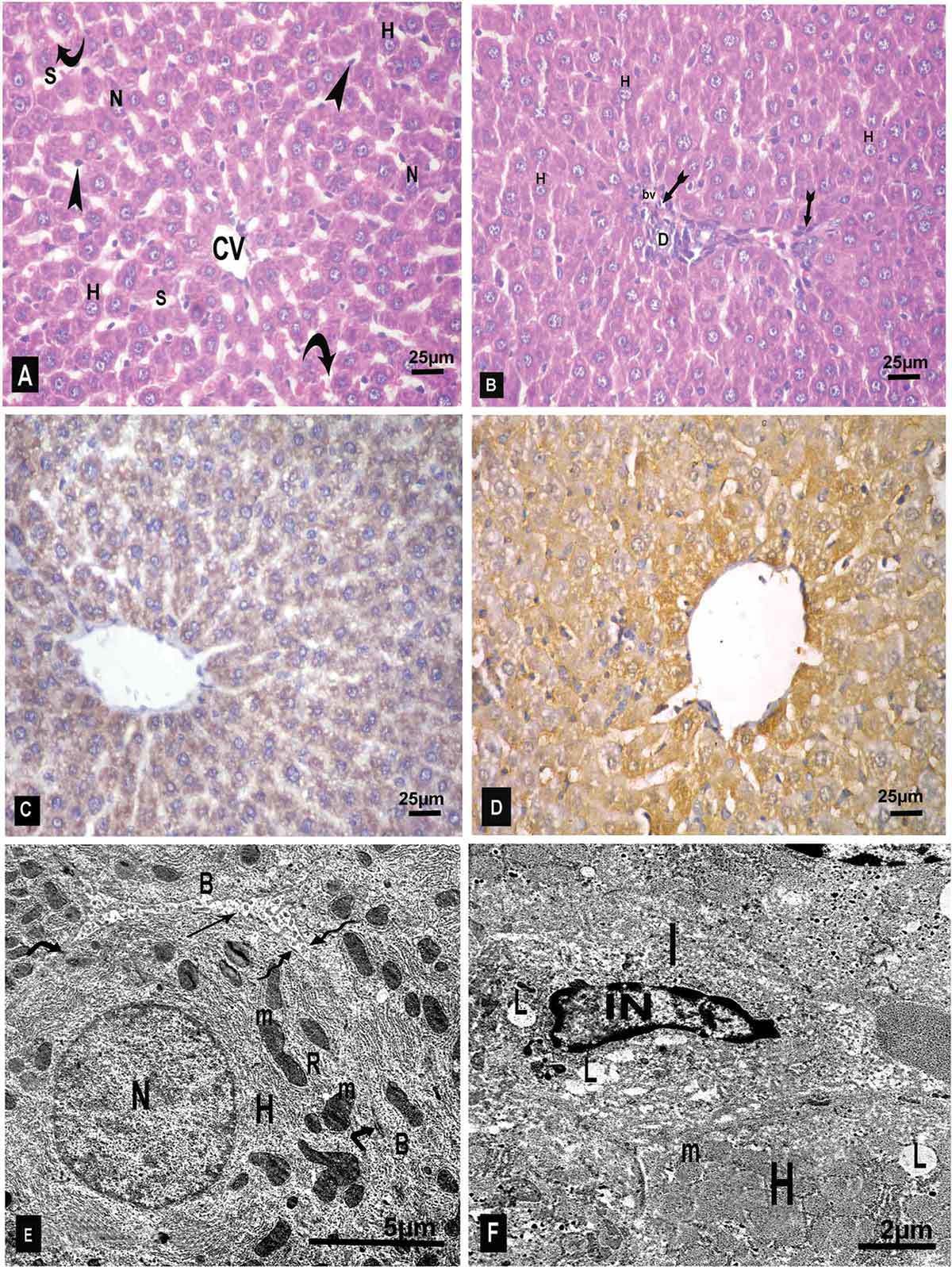
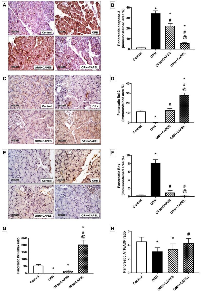
- Effect of administering caffeic acid phenethyl ester (CAPE)-optimal liposomal formulation (CAPEL) on pancreatic apoptosis and energy status in rats with ornithine-induced acute pancreatitis. ( A ) Representative photomicrographs of the immunohistochemical evaluation of pancreatic cleaved caspase-3 protein expression in the different study groups (x400 magnification). ( B ) Quantification of cleaved caspase-3 immunostaining in pancreatic tissue. ( C ) Representative photomicrographs of the immunohistochemical evaluation of pancreatic Bcl-2 protein expression in the different study groups (x400 magnification). ( D ) Quantification of Bcl-2 immunostaining in pancreatic tissue. ( E ) Representative photomicrographs of the immunohistochemical evaluation of pancreatic Bax protein expression in the different study groups (x400 magnification). ( F ) Quantification of Bax immunostaining in pancreatic tissue. Values in ( B , D , F ) are the mean +- S.D. of the area percentage of cleaved caspase-3, Bcl-2 and Bax immunostaining, respectively, to the total area of the microscopic field across six non-overlapping fields/section ( n = 8). ( G ) Pancreatic Bcl-2/Bax ratio. ( H ) Pancreatic ATP/ADP ratio. Data are expressed as mean +- S.D. ( n = 8). *, # and @ indicate significant difference at p < 0.05 from control, ORN and ORN+CAPES groups, respectively (one-way ANOVA followed by Tukey's multiple comparisons test). ORN, ornithine; CAPES, free caffeic acid phenethyl ester (CAPE) suspension;
- Submitted by
- Invitrogen Antibodies (provider)
- Main image
- Experimental details
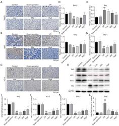
- Figure 2 AS-IV treatment relieved I/R-induced apoptosis and regulated the Nrf2/HO-1 pathway in rat kidney tissues. (A) Apoptosis in the renal tissues of AKI rats as detected by TUNEL staining. Magnification x400. (B,C) Nrf2 and HO-1 were detected by IHC. Magnification x400. Bcl-2, Bax, Nrf2, and HO-1 expression were evaluated by qRT-PCR (D-G) and western blotting (H-L). # P
