Explore
 Validate
 Learn
Learn Immunocytochemistry
Antibody data
- Antibody Data
- Antigen structure
- References [5]
- Comments [0]
- Validations
- Immunocytochemistry [2]
- Immunohistochemistry [1]
- Other assay [1]
Submit
Validation data
Reference
Comment
Report error
- Product number
- MA5-11579 - Provider product page

- Provider
- Invitrogen Antibodies
- Product name
- CA125 Monoclonal Antibody (Ov185:1)
- Antibody type
- Monoclonal
- Antigen
- Purifed from natural sources
- Description
- MA5-11579 targets CA-125 in IHC (P) applications and shows reactivity with Human samples. The MA5-11579 immunogen is partially purified mucin fraction from a pool of cancer tissue from patients with epithelial ovarian cancer.
- Reactivity
- Human
- Host
- Mouse
- Isotype
- IgG
- Antibody clone number
- Ov185:1
- Vial size
- 500 μL
- Concentration
- Conc. Not Determined
- Storage
- 4°C
Submitted references Development of a near infrared protein nanoprobe targeting Thomsen-Friedenreich antigen for intraoperative detection of submillimeter nodules in an ovarian peritoneal carcinomatosis mouse model.
Dual use of a single Wilms' tumor 1 immunohistochemistry in evaluation of ovarian tumors: a preliminary study of 20 cases.
Dual use of a single Wilms' tumor 1 immunohistochemistry in evaluation of ovarian tumors: a preliminary study of 20 cases.
Multiple initial culture conditions enhance the establishment of cell lines from primary ovarian cancer specimens.
Prognostic significance of CA 125, CD44, and epithelial membrane antigen in renal cell carcinoma.
Coustets M, Ladurantie C, Bellard E, Prat M, Rols MP, Ecochard V, Ferron G, Chabot S, Golzio M, Paquereau L
Biomaterials 2020 May;241:119908
Biomaterials 2020 May;241:119908
Dual use of a single Wilms' tumor 1 immunohistochemistry in evaluation of ovarian tumors: a preliminary study of 20 cases.
Hsiao YH, Siddiqui S, Man YG
Journal of Cancer 2010 Jul 13;1:93-7
Journal of Cancer 2010 Jul 13;1:93-7
Dual use of a single Wilms' tumor 1 immunohistochemistry in evaluation of ovarian tumors: a preliminary study of 20 cases.
Hsiao YH, Siddiqui S, Man YG
Journal of Cancer 2010 Jul 13;1:93-7
Journal of Cancer 2010 Jul 13;1:93-7
Multiple initial culture conditions enhance the establishment of cell lines from primary ovarian cancer specimens.
Bertozzi CC, Chang CY, Jairaj S, Shan X, Huang J, Weber BL, Chu CS, Carroll RG
In vitro cellular & developmental biology. Animal 2006 Mar-Apr;42(3-4):58-62
In vitro cellular & developmental biology. Animal 2006 Mar-Apr;42(3-4):58-62
Prognostic significance of CA 125, CD44, and epithelial membrane antigen in renal cell carcinoma.
Bamias A, Chorti M, Deliveliotis C, Trakas N, Skolarikos A, Protogerou B, Legaki S, Tsakalou G, Tamvakis N, Dimopoulos MA
Urology 2003 Aug;62(2):368-73
Urology 2003 Aug;62(2):368-73
No comments: Submit comment
Supportive validation
- Submitted by
- Invitrogen Antibodies (provider)
- Main image

- Experimental details
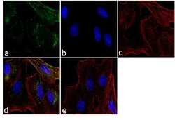
- Immunofluorescence analysis of CA-125 / MUC16 was performed using 70% confluent log phase SK-OV-3 cells. The cells were fixed with 4% paraformaldehyde for 10 minutes, permeabilized with 0.1% Triton™ X-100 for 10 minutes, and blocked with 1% BSA for 1 hour at room temperature. The cells were labeled with CA-125 / MUC16 (Ov185:1) Mouse Monoclonal Antibody (Product # MA5-11579) at 1:250 dilution in 0.1% BSA and incubated for 3 hours at room temperature and then labeled with Goat anti-Mouse IgG (H+L) Superclonal™ Secondary Antibody, Alexa Fluor® 488 conjugate (Product # A28175) a dilution of 1:2000 for 45 minutes at room temperature (Panel a: green). Nuclei (Panel b: blue) were stained with SlowFade® Gold Antifade Mountant with DAPI (Product # S36938). F-actin (Panel c: red) was stained with Rhodamine Phalloidin (Product # R415, 1:300). Panel d represents the merged image showing cytoplasmic localization. Panel e shows the no primary antibody control. The images were captured at 60X magnification.

- Submitted by
- Invitrogen Antibodies (provider)
- Main image
- Experimental details
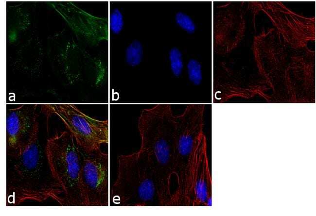
- Immunofluorescence analysis of CA-125 / MUC16 was performed using 70% confluent log phase SK-OV-3 cells. The cells were fixed with 4% paraformaldehyde for 10 minutes, permeabilized with 0.1% Triton™ X-100 for 10 minutes, and blocked with 1% BSA for 1 hour at room temperature. The cells were labeled with CA-125 / MUC16 (Ov185:1) Mouse Monoclonal Antibody (Product # MA5-11579) at 1:250 dilution in 0.1% BSA and incubated for 3 hours at room temperature and then labeled with Goat anti-Mouse IgG (H+L) Superclonal™ Secondary Antibody, Alexa Fluor® 488 conjugate (Product # A28175) a dilution of 1:2000 for 45 minutes at room temperature (Panel a: green). Nuclei (Panel b: blue) were stained with SlowFade® Gold Antifade Mountant with DAPI (Product # S36938). F-actin (Panel c: red) was stained with Rhodamine Phalloidin (Product # R415, 1:300). Panel d represents the merged image showing cytoplasmic localization. Panel e shows the no primary antibody control. The images were captured at 60X magnification.
Supportive validation
- Submitted by
- Invitrogen Antibodies (provider)
- Main image

- Experimental details
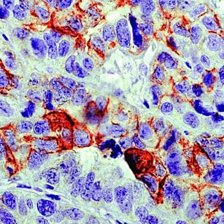
- Formalin-fixed, paraffin-embedded human ovarian carcinoma stained with CA125 antibody using peroxidase-conjugate and AEC chromogen. Note cell membrane staining of tumor cells.
Supportive validation
- Submitted by
- Invitrogen Antibodies (provider)
- Main image
- Experimental details
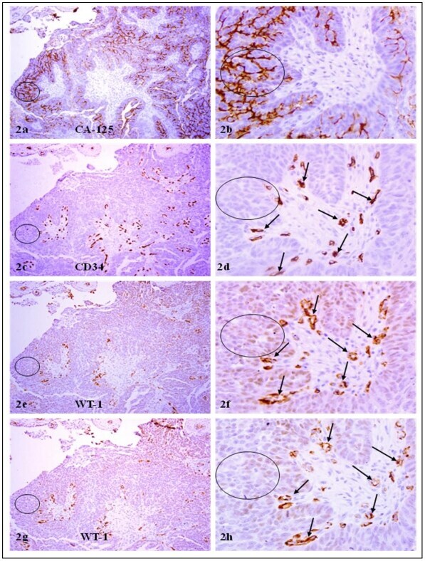
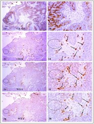
- Figure 2 CA-125 or CD 34 positive cells co-express WT-1. A set of 4-consecutive sections were immunostained for CA-125, CD34, and WT-1. Circles identify tumor cell clusters with WT-1 and CA-125 expression. Arrows identify small blood vessels. Note that a vast majority of CA-125 or CD34 positive tumor or endothelial cells co-express WT-1, and that WT-1 expression was consistent in different sections. a, c, e, and g: 100X. b, d, f, and h: a higher (300X) magnification of a, c, e, and g, respectively.