Explore
 Validate
 Learn
Learn Western blot
Antibody data
- Antibody Data
- Antigen structure
- References [6]
- Comments [0]
- Validations
- Western blot [1]
- Immunohistochemistry [2]
- Other assay [2]
Submit
Validation data
Reference
Comment
Report error
- Product number
- PA1-37282 - Provider product page

- Provider
- Invitrogen Antibodies
- Product name
- CD3e Polyclonal Antibody
- Antibody type
- Polyclonal
- Antigen
- Synthetic peptide
- Description
- Heat-mediated antigen retrieval is recommended prior to staining, using a 10mM citrate buffer, pH 6.0, for 10 minutes followed by cooling at room temperature for 20 min. Following antigen retrieval, incubate samples with primary antibody for 10 min at room temperature. A suggested positive control is tonsil tissue.
- Reactivity
- Human
- Host
- Rabbit
- Isotype
- IgG
- Vial size
- 1 mL
- Concentration
- 1.06 mg/mL
- Storage
- -20° C, Avoid Freeze/Thaw Cycles
Submitted references Investigation of CD26, a potential SARS-CoV-2 receptor, as a biomarker of age and pathology.
OTUB1 inhibits CNS autoimmunity by preventing IFN-γ-induced hyperactivation of astrocytes.
Stem cells from human exfoliated deciduous teeth correct the immune imbalance of allergic rhinitis via Treg cells in vivo and in vitro.
Tumor-immune profiling of murine syngeneic tumor models as a framework to guide mechanistic studies and predict therapy response in distinct tumor microenvironments.
Highly Proliferative α-Cell-Related Islet Endocrine Cells in Human Pancreata.
Extreme Beta-Cell Deficiency in Pancreata of Dogs with Canine Diabetes.
Raha AA, Chakraborty S, Henderson J, Mukaetova-Ladinska E, Zaman S, Trowsdale J, Raha-Chowdhury R
Bioscience reports 2020 Dec 23;40(12)
Bioscience reports 2020 Dec 23;40(12)
OTUB1 inhibits CNS autoimmunity by preventing IFN-γ-induced hyperactivation of astrocytes.
Wang X, Mulas F, Yi W, Brunn A, Nishanth G, Just S, Waisman A, Brück W, Deckert M, Schlüter D
The EMBO journal 2019 May 15;38(10)
The EMBO journal 2019 May 15;38(10)
Stem cells from human exfoliated deciduous teeth correct the immune imbalance of allergic rhinitis via Treg cells in vivo and in vitro.
Dai YY, Ni SY, Ma K, Ma YS, Wang ZS, Zhao XL
Stem cell research & therapy 2019 Jan 22;10(1):39
Stem cell research & therapy 2019 Jan 22;10(1):39
Tumor-immune profiling of murine syngeneic tumor models as a framework to guide mechanistic studies and predict therapy response in distinct tumor microenvironments.
Yu JW, Bhattacharya S, Yanamandra N, Kilian D, Shi H, Yadavilli S, Katlinskaya Y, Kaczynski H, Conner M, Benson W, Hahn A, Seestaller-Wehr L, Bi M, Vitali NJ, Tsvetkov L, Halsey W, Hughes A, Traini C, Zhou H, Jing J, Lee T, Figueroa DJ, Brett S, Hopson CB, Smothers JF, Hoos A, Srinivasan R
PloS one 2018;13(11):e0206223
PloS one 2018;13(11):e0206223
Highly Proliferative α-Cell-Related Islet Endocrine Cells in Human Pancreata.
Lam CJ, Cox AR, Jacobson DR, Rankin MM, Kushner JA
Diabetes 2018 Apr;67(4):674-686
Diabetes 2018 Apr;67(4):674-686
Extreme Beta-Cell Deficiency in Pancreata of Dogs with Canine Diabetes.
Shields EJ, Lam CJ, Cox AR, Rankin MM, Van Winkle TJ, Hess RS, Kushner JA
PloS one 2015;10(6):e0129809
PloS one 2015;10(6):e0129809
No comments: Submit comment
Supportive validation
- Submitted by
- Invitrogen Antibodies (provider)
- Main image
- Experimental details
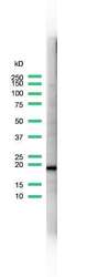
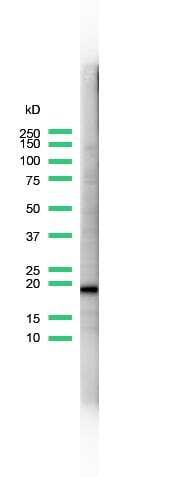
- Western blot analysis of Jurkat Cells using anti-CD3 Polyclonal Antibody (Product # PA5-32318). The recommened dilution for this antibody in western blot applications is 1:25.
Supportive validation
- Submitted by
- Invitrogen Antibodies (provider)
- Main image

- Experimental details
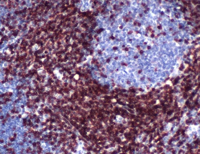
- Immunohistochemical analysis of CD3e using a polyclonal antibody (Product # PA1-37282).
- Submitted by
- Invitrogen Antibodies (provider)
- Main image

- Experimental details
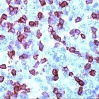
- Immunohistochemical analysis of CD3 using anti-CD3 Polyclonal Antibody (Product # PA5-32318) in Tonsil Tissue. The recommened dilution for this antibody in immunohistochemistry applications is 1:200.
Supportive validation
- Submitted by
- Invitrogen Antibodies (provider)
- Main image

- Experimental details
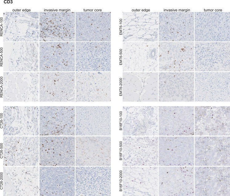
- Fig 9 CD3 + cells are confined predominantly to the invasive margin in untreated tumors. IHC was performed on fixed and paraffin embedded tumor samples across the different models and across all tumor sizes. Five mice per model at each tumor size were used for this analysis. A representative image for each is shown.
- Submitted by
- Invitrogen Antibodies (provider)
- Main image

- Experimental details
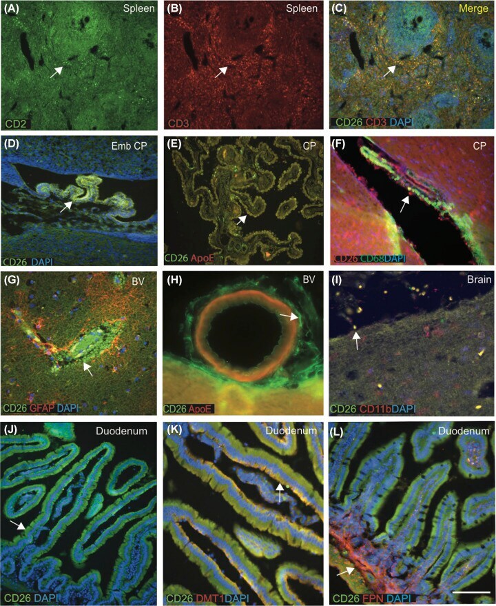
- Figure 2 CD26 protein is expressed in the CP epithelial cells, meningeal blood vessels and bind with divalent metal proteins in the duodenum Double IFC staining was performed in the human spleen using mouse mAb anti-CD26 (Green) and rabbit polyclonal (pAb) anti-CD3 (red), and DAPI for nuclei (blue) ( A - C ). CD26 immunoreactivity was seen in the human spleen in white pulp, very close to the central canal and in T-cell area surrounding the lymphoid follicles (A,B). CD26 and CD3 (a marker expressed in T cells) colocalised in the outer darker region of the germinal centre containing large- and medium-sized T lymphocytes and both proteins colocalised (C). Embryonic mouse CP was stained with CD26 (green) and DAPI (blue) and imaged using confocal microscopy. Very high CD26 expression was present in the mouse embryonic CP epithelium ( D ). Human normal brain section particularly CP was stained with CD26 (green) and ApoE (red), both proteins were present in the CP epithelial membrane and colocalised in the macrophages ( E ). A mouse brain section from lateral ventricle stained with CD26 (red) and a macrophages marker CD68 (green), CD26-positive macrophages were visible at the wall of ventricle and in the CP, both proteins colocalised ( F ). An ARD brain section from cortex, close to a blood vessel was stained with CD26 (green) and astrocyte marker (GFAP, red), showed that CD26 protein carried by RBCs and macrophages entering through damaged blood vessels (BVs), surrounded by GFAP-po