 Explore
 Validate
 Learn
Learn Western blot
 Immunocytochemistry
Antibody data
- Antibody Data
- Antigen structure
- References [2]
- Comments [0]
- Validations
- Immunocytochemistry [3]
- Immunohistochemistry [1]
- Other assay [1]
Submit
Validation data
Reference
Comment
Report error
- Product number
- PA5-20202 - Provider product page

- Provider
- Invitrogen Antibodies
- Product name
- TLR9 Polyclonal Antibody
- Antibody type
- Polyclonal
- Antigen
- Synthetic peptide
- Description
- A suggested positive control is mouse spleen tissue lysate. PA5-20202 can be used with blocking peptide PEP-0330.
- Reactivity
- Human, Mouse
- Host
- Rabbit
- Isotype
- IgG
- Vial size
- 100 μg
- Concentration
- 1 mg/mL
- Storage
- Maintain refrigerated at 2-8°C for up to 3 months. For long term storage store at -20°C
Submitted references Toll-Like Receptor 9 Expression Levels in Breast Carcinoma Correlate with Improved Overall Survival in Patients Treated with Neoadjuvant Chemotherapy and Could Serve as a Prognostic Marker.
Influenza-induced immune suppression to methicillin-resistant Staphylococcus aureus is mediated by TLR9.
Singh A, Bandyopadhyay A, Mukherjee N, Basu A
Genetic testing and molecular biomarkers 2021 Jan;25(1):12-19
Genetic testing and molecular biomarkers 2021 Jan;25(1):12-19
Influenza-induced immune suppression to methicillin-resistant Staphylococcus aureus is mediated by TLR9.
Martínez-Colón GJ, Warheit-Niemi H, Gurczynski SJ, Taylor QM, Wilke CA, Podsiad AB, Crespo J, Bhan U, Moore BB
PLoS pathogens 2019 Jan;15(1):e1007560
PLoS pathogens 2019 Jan;15(1):e1007560
No comments: Submit comment
Supportive validation
- Submitted by
- Invitrogen Antibodies (provider)
- Main image
- Experimental details
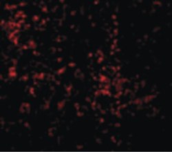
- Immunofluorescent analysis of mouse spleen cells using a CD289/TLR9 polyclonal antibody (Product # PA5-20202) at a 10 µg/mL dilution.

- Submitted by
- Invitrogen Antibodies (provider)
- Main image

- Experimental details
- Immunofluorescence of TLR9 in mouse spleen cells with TLR9 Polyclonal Antibody (Product # PA5-20202) at 10 µg/mL.
- Submitted by
- Invitrogen Antibodies (provider)
- Main image

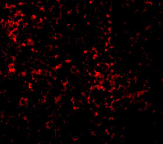
- Experimental details
- Immunofluorescence of TLR9 in mouse spleen cells with TLR9 Polyclonal Antibody (Product # PA5-20202) at 10 µg/mL.

Supportive validation
- Submitted by
- Invitrogen Antibodies (provider)
- Main image
- Experimental details
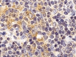
- Immunohistochemistry of TLR9 in mouse spleen cells with TLR9 Polyclonal Antibody (Product # PA5-20202) at 2 µg/mL.

Supportive validation
- Submitted by
- Invitrogen Antibodies (provider)
- Main image
- Experimental details
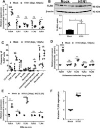
- Fig 1 TLR9 overexpression in lung immune cells post-IAV infection. (A) Relative gene expression of TLRs (9, 7, 3, 2, 4) by RTqPCR and (B) western blotting of TLR9 and beta-actin. RNA and protein were isolated from lung leukocytes post-collagenase digestion in mice infected with 100 PFUs of H1N1 (PR8) for 5 days or placebo (PBS). beta-actin was used to normalize RNA in samples. (C) Frequency of TLR9 + cells measured by flow cytometry in lung immune cells post-collagenase digestion. Gating was as follows: CD4 + T cells (CD45 + ,CD90.2 + , CD3 + , CD4 + ), CD8 + T cells (CD45 + ,CD90.2 + , CD3 + , CD4 - , CD8 + ), natural killer cells (CD45 + ,CD90.2 +, NKp46 + ), B cells (CD45 + ,CD90.2 - , CD19 + ), neutrophils (CD45 + , CD11b + , LY6G + ), interstitial macrophages (CD45 + , CD64 + , CD11b + , F4/80 + ), alveolar macrophages (CD45 + , CD64 + , CD11C + , Siglec F + ) and dendritic cells (CD45 + , CD64 - , CD11c + , MCHII high ). Staining for TLR9 was performed on cells which were fixed and permeabilized. (D) Relative expression of TLRs (9, 7, 3, 2, 4) after lung macrophage isolation by adherence selection from PBS or IAV-infected mice. (E) Relative expression of TLRs (9, 7, 3, 2, 4) in alveolar macrophages infected or not ex-vivo with H1N1 (MOI:0.01) for 24 hours. (F) BMDMs from Balb/c mice infected with H1N1 at MOI:0.01 and measured for TLR9 mRNA after 48h. Statistics are student T test between comparative groups. *P
