Explore
 Validate
 Learn
Learn Western blot
Antibody data
- Antibody Data
- Antigen structure
- References [4]
- Comments [0]
- Validations
- Western blot [4]
- Immunocytochemistry [2]
- Other assay [2]
Submit
Validation data
Reference
Comment
Report error
- Product number
- PA5-20203 - Provider product page

- Provider
- Invitrogen Antibodies
- Product name
- TLR9 Polyclonal Antibody
- Antibody type
- Polyclonal
- Antigen
- Synthetic peptide
- Description
- A suggested positive control is Jurkat cell lysate. PA5-20203 can be used with blocking peptide PEP-0346.
- Reactivity
- Human, Mouse, Rat
- Host
- Rabbit
- Isotype
- IgG
- Vial size
- 100 µg
- Concentration
- 1 mg/mL
- Storage
- 4° C
Submitted references Chronic Inhibition of Toll-Like Receptor 9 Ameliorates Pulmonary Hypertension in Rats.
Munc13-4 Is a Rab11-binding Protein That Regulates Rab11-positive Vesicle Trafficking and Docking at the Plasma Membrane.
Munc13-4 interacts with syntaxin 7 and regulates late endosomal maturation, endosomal signaling, and TLR9-initiated cellular responses.
Phagocytosis-dependent activation of a TLR9-BTK-calcineurin-NFAT pathway co-ordinates innate immunity to Aspergillus fumigatus.
Ishikawa T, Abe K, Takana-Ishikawa M, Yoshida K, Watanabe T, Imakiire S, Hosokawa K, Hirano M, Hirano K, Tsutsui H
Journal of the American Heart Association 2021 Apr 6;10(7):e019247
Journal of the American Heart Association 2021 Apr 6;10(7):e019247
Munc13-4 Is a Rab11-binding Protein That Regulates Rab11-positive Vesicle Trafficking and Docking at the Plasma Membrane.
Johnson JL, He J, Ramadass M, Pestonjamasp K, Kiosses WB, Zhang J, Catz SD
The Journal of biological chemistry 2016 Feb 12;291(7):3423-38
The Journal of biological chemistry 2016 Feb 12;291(7):3423-38
Munc13-4 interacts with syntaxin 7 and regulates late endosomal maturation, endosomal signaling, and TLR9-initiated cellular responses.
He J, Johnson JL, Monfregola J, Ramadass M, Pestonjamasp K, Napolitano G, Zhang J, Catz SD
Molecular biology of the cell 2016 Feb 1;27(3):572-87
Molecular biology of the cell 2016 Feb 1;27(3):572-87
Phagocytosis-dependent activation of a TLR9-BTK-calcineurin-NFAT pathway co-ordinates innate immunity to Aspergillus fumigatus.
Herbst S, Shah A, Mazon Moya M, Marzola V, Jensen B, Reed A, Birrell MA, Saijo S, Mostowy S, Shaunak S, Armstrong-James D
EMBO molecular medicine 2015 Mar;7(3):240-58
EMBO molecular medicine 2015 Mar;7(3):240-58
No comments: Submit comment
Supportive validation
- Submitted by
- Invitrogen Antibodies (provider)
- Main image

- Experimental details
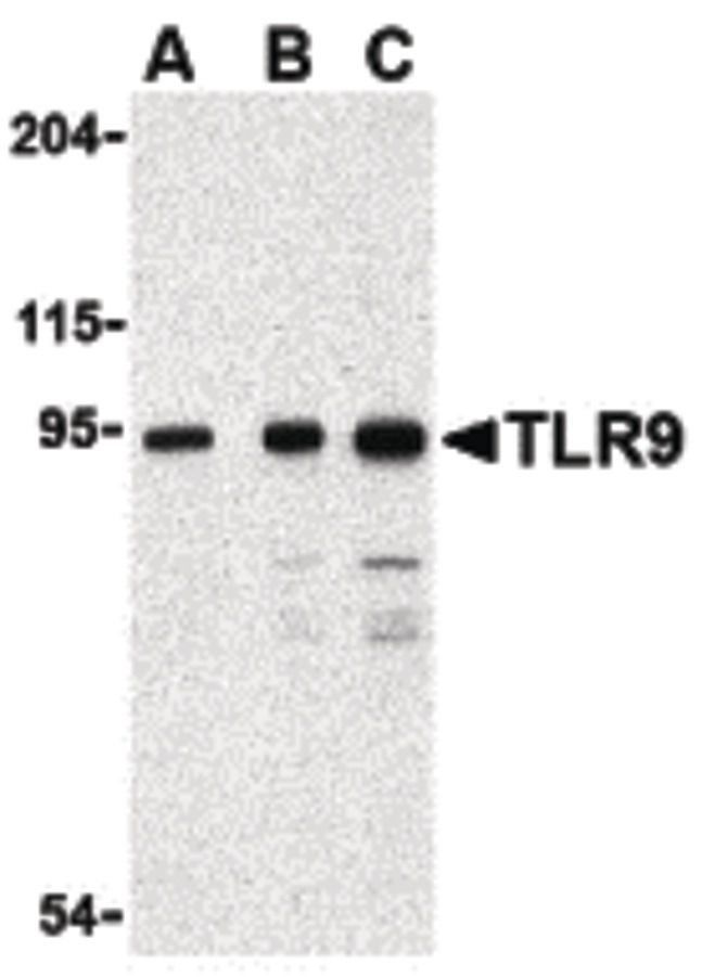
- Western blot analysis of Jurkat cell lysate using a CD289/TLR9 polyclonal antibody (Product # PA5-20203) at (A) 0.5, (B) 1 and (C) 2 µg/mL.
- Submitted by
- Invitrogen Antibodies (provider)
- Main image
- Experimental details
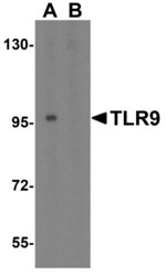
- Western Blot analysis of TLR9 in Jurkat cell lysate with TLR9 Polyclonal Antibody (Product # PA5-20203) at 1 µg/mL in (A) the absence and (B) the presence of blocking peptide

- Submitted by
- Invitrogen Antibodies (provider)
- Main image
- Experimental details
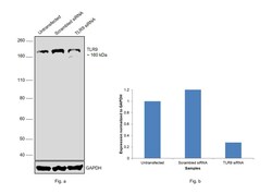
- Knockdown of Toll-like receptor 9 was achieved by transfecting SH-SY5Y with TLR9 specific siRNAs (Silencer® select Product # s28872, s28874). Western blot analysis (Fig. a) was performed using whole cell extracts from the TLR9 knockdown cells (lane 3), non-targeting scrambled siRNA transfected cells (lane 2) and untransfected cells (lane 1). The blot was probed with TLR9 Polyclonal Antibody (Product # PA5-20203, 1.0 µg/mL dilution) and Goat anti-Rabbit IgG (H+L) Superclonal™ Recombinant Secondary Antibody, HRP (Product # A27036, 1:10000 dilution). Densitometric analysis of this western blot is shown in histogram (Fig. b). Decrease in signal upon siRNA mediated knock down confirms that antibody is specific to Toll-like receptor 9.

- Submitted by
- Invitrogen Antibodies (provider)
- Main image
- Experimental details
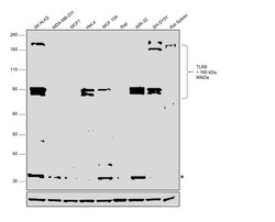
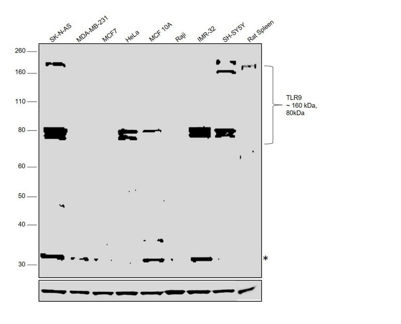
- Western blot was performed using Anti-TLR9 Polyclonal Antibody (Product # PA5-20203) and a 160 kDa full length and 80 kDa cleaved form corresponding to Toll-like receptor 9 was observed across cell lines and tissues tested. Whole cell extracts (40 µg lysate) of SK-N-AS (Lane 1), MDA-MB-231 (Lane 2), MCF7 (Lane 3), HeLa (Lane 4), MCF 10A (Lane 5), Raji (Lane 6), IMR-32 (Lane 7), SH-SY5Y (Lane 8) and tissue extracts of Rat Spleen (Lane 9) were electrophoresed using NuPAGE™ 4-12% Bis-Tris Protein Gel (Product # NP0321BOX). Resolved proteins were then transferred onto a Nitrocellulose membrane (Product # IB23001) by iBlot® 2 Dry Blotting System (Product # IB21001). The blot was probed with the primary antibody (1.0 µg/mL dilution) and detected by chemiluminescence with Goat anti-Rabbit IgG (H+L) Superclonal™ Recombinant Secondary Antibody, HRP (Product # A27036, 1:10000 dilution) using the iBright FL 1000 (Product # A32752). Chemiluminescent detection was performed using Novex® ECL Chemiluminescent Substrate Reagent Kit (Product # WP20005).
Supportive validation
- Submitted by
- Invitrogen Antibodies (provider)
- Main image

- Experimental details
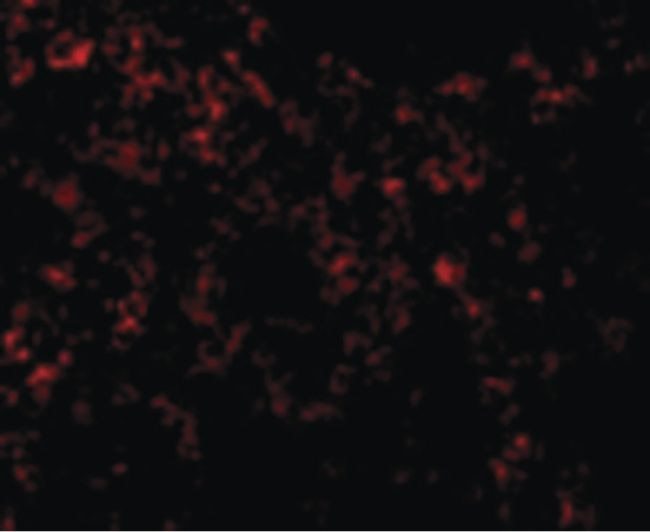
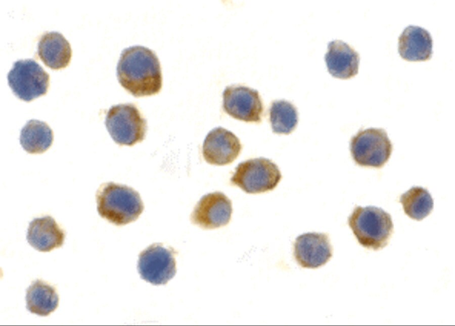
- Immunofluorescent analysis of mouse spleen cells using a CD289/TLR9 polyclonal antibody (Product # PA5-20203) at a 10 µg/mL dilution.
- Submitted by
- Invitrogen Antibodies (provider)
- Main image
- Experimental details
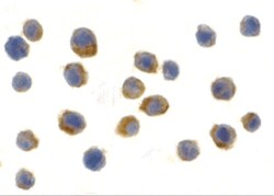
- Immunocytochemistry of TLR9 in Jurkat cells with TLR9 Polyclonal Antibody (Product # PA5-20203) at 2 µg/mL.
Supportive validation
- Submitted by
- Invitrogen Antibodies (provider)
- Main image
- Experimental details
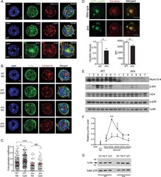
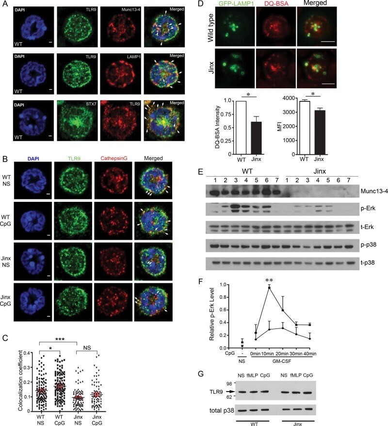
- FIGURE 6: Munc13-4 is required for CpG-ODN-induced endosomal maturation and TLR9-dependent signaling. (A) Confocal microscopy analysis of the distribution of TLR9 with Munc13-4, LAMP1, and STX7 in WT neutrophils. Cell nuclei are visualized with DAPI staining (blue). Scale bar, 1 mum. (B) Confocal microscopy analysis of the distribution of TLR9 with the protease cathepsin G in WT and Munc13-4-KO ( Jinx ) neutrophils. NS, no stimulation; CpG, cells were primed with GM-CSF and treated with CPG-ODN. Scale bar, 1 mum. (C) Quantification analysis showing the percentage of TLR9-positive vesicles that contain cathepsin G. In these assays, 109, 113, 131, and 137 cells were analyzed for the groups WT NS, WT+CpG, Jinx NS, and Jinx +CpG, respectively. Results are expressed mean +- SEM. * p < 0.05, *** p < 0.0001. (D) Top, pTIRFM images of WT and Jinx neutrophils expressing GFP-LAMP1 (green) with internalized DQ-BSA (red), a fluorescent probe whose intensity increases upon degradation. The fluorescence intensity of the probe was analyzed from confocal images using ImageJ (bottom left) or by flow cytometry (bottom right). Results are mean +- SEMs. For the quantification of confocal images, 342 WT and 344 Jinx neutrophils were analyzed. For flow cytometry, n = 3 for both WT and Jinx. * p < 0.05. (E) Western blot analysis of neutrophil signaling in response to TLR9 activation. WT and Munc13-4-KO ( Jinx ) neutrophils were treated as follows: 1, untreated; 2, GM-CSF 1.5 h; 3, GM-CSF 1.5 h + Cp
- Submitted by
- Invitrogen Antibodies (provider)
- Main image
- Experimental details
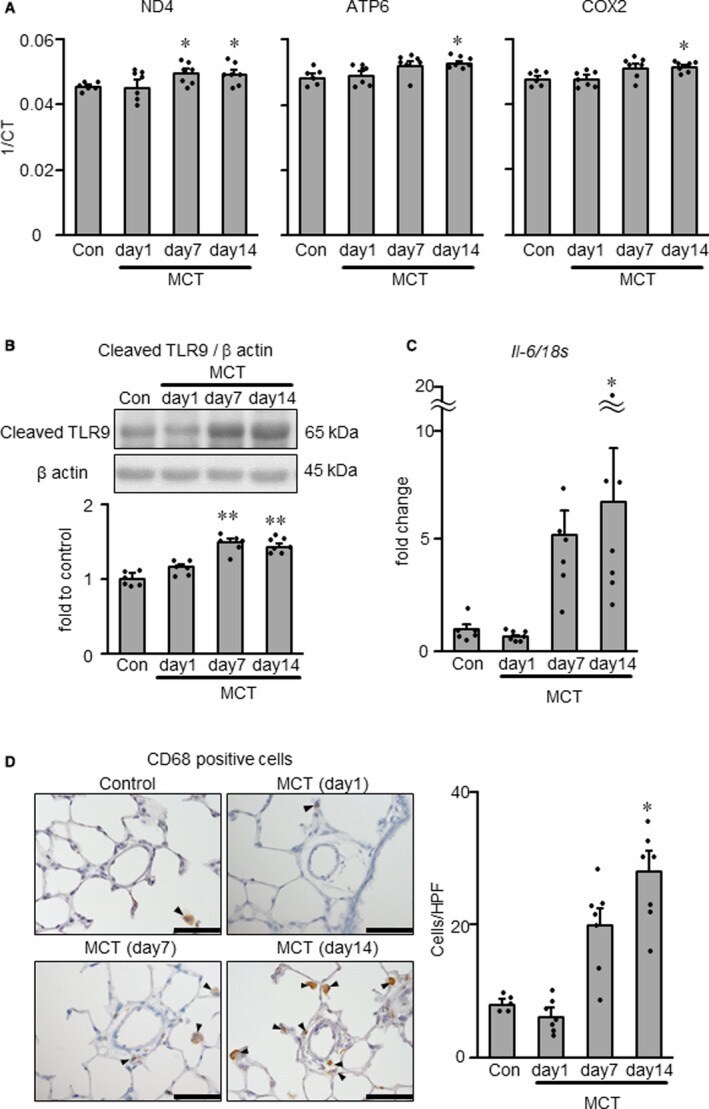
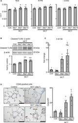
- Figure 2 Quantification of mitochondrial DNA genes, TLR9 activation, Il-6 and macrophage accumulation in normal control and MCT-exposed rats. A , NADH dehydrogenase subunit 4 (ND4, left), ATP synthase 6 (ATP6, middle) and cytochrome C oxidase subunit II (COX2, right) in plasma of normal and MCT-exposed rats at days 1, 7, and 14 after MCT injection, as measured by real-time PCR. 1/CT denotes the reciprocal of the count where the sequence is detected, and is a direct function of gene concentration. B , Protein expression level of cleaved TLR9 in whole lung measured by Western blotting. C , Il-6 mRNA expression level in whole lung measured by RT-PCR. D , Representative photomicrographs (left) and counts (right) of CD68-positive macrophages (arrowheads) in the lung. Scale bars indicate 50 mum. Con indicates normal rats injected with PBS; and MCT, monocrotaline. N=5-7. * P