 Explore
 Validate
 Learn
Learn Immunohistochemistry
Antibody data
- Antibody Data
- Antigen structure
- References [1]
- Comments [0]
- Validations
- Immunohistochemistry [1]
- Flow cytometry [1]
Submit
Validation data
Reference
Comment
Report error
- Product number
- AF3658 - Provider product page

- Provider
- R&D Systems
- Product name
- Human TLR9 Antibody
- Antibody type
- Polyclonal
- Description
- Immunogen affinity purified. Detects human TLR9 in direct ELISAs. In direct ELISAs, approximately 75% cross-reactivity with recombinant mouse TLR9 is observed, and less than 1% cross-reactivity with recombinant human (rh) TLR1, rhTLR2, rhTLR3, rhTLR4, rhTLR5, rhTLR7, rhTLR8, and rhTLR10 is observed.
- Reactivity
- Human
- Host
- Sheep
- Conjugate
- Unconjugated
- Antigen sequence
Q9NR96- Isotype
- IgG
- Vial size
- 100 ug
- Concentration
- LYOPH
- Storage
- Use a manual defrost freezer and avoid repeated freeze-thaw cycles. 12 months from date of receipt, -20 to -70 °C as supplied. 1 month, 2 to 8 °C under sterile conditions after reconstitution. 6 months, -20 to -70 °C under sterile conditions after reconstitution.
Submitted references FcγRIIIa Signaling Modulates Endosomal TLR Responses in Human CD4(+) T Cells.
Chauhan AK
Journal of immunology (Baltimore, Md. : 1950) 2017 Jun 15;198(12):4596-4606
Journal of immunology (Baltimore, Md. : 1950) 2017 Jun 15;198(12):4596-4606
No comments: Submit comment
Supportive validation
- Submitted by
- R&D Systems (provider)
- Main image

- Experimental details
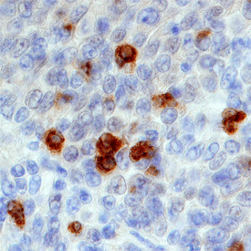
- TLR9 in Human Tonsil. TLR9 was detected in immersion fixed paraffin-embedded sections of human tonsil using Sheep Anti-Human TLR9 Antigen Affinity-purified Polyclonal Antibody (Catalog # AF3658) at 1 µg/mL overnight at 4 °C. Tissue was stained using the Anti-Sheep HRP-DAB Cell & Tissue Staining Kit (brown; Catalog # CTS019) and counterstained with hematoxylin (blue). Specific staining was localized to membranes of lymphocytes. View our protocol for Chromogenic IHC Staining of Paraffin-embedded Tissue Sections.
Supportive validation
- Submitted by
- R&D Systems (provider)
- Main image
- Experimental details
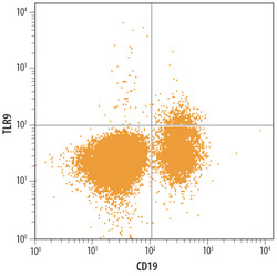
- Detection of TLR9 in Human PBMC lymphocytes by Flow Cytometry. Human PBMC lymphocytes were stained with Sheep Anti-Human TLR9 Antigen Affinity-purified Polyclonal Antibody (Catalog # AF3658) followed by NorthernLights™ 637-conjugated Anti-Sheep IgG Secondary Antibody (Catalog # NL011) and Mouse Anti-Human CD19 PE-conjugated Monoclonal Antibody (Catalog # FAB4867P). Quadrant markers were set based on control antibody staining (Catalog # 5-001-A).
