Explore
 Validate
 Learn
Learn Western blot
 Immunohistochemistry
Antibody data
- Antibody Data
- Antigen structure
- References [1]
- Comments [0]
- Validations
- Immunohistochemistry [1]
Submit
Validation data
Reference
Comment
Report error
- Product number
- HPA002859 - Provider product page

- Provider
- Atlas Antibodies
- Proper citation
- Atlas Antibodies Cat#HPA002859, RRID:AB_1079672
- Product name
- Anti-PRAF2
- Antibody type
- Polyclonal
- Description
- Polyclonal Antibody against Human PRAF2, Gene description: PRA1 domain family, member 2, Alternative Gene Names: JM4, Validated applications: WB, IHC, Uniprot ID: O60831, Storage: Store at +4°C for short term storage. Long time storage is recommended at -20°C.
- Reactivity
- Human, Rat
- Host
- Rabbit
- Conjugate
- Unconjugated
- Isotype
- IgG
- Vial size
- 100 µl
- Concentration
- 0.1 mg/ml
- Storage
- Store at +4°C for short term storage. Long time storage is recommended at -20°C.
- Handling
- The antibody solution should be gently mixed before use.
Submitted references Expression profile of PRAF2 in the human brain and enrichment in synaptic vesicles
Koomoa D, Go R, Wester K, Bachmann A
Neuroscience Letters 2008;436(2):171-176
Neuroscience Letters 2008;436(2):171-176
No comments: Submit comment
Supportive validation
- Submitted by
- Atlas Antibodies (provider)
- Enhanced method
- Orthogonal validation
- Main image
- Experimental details
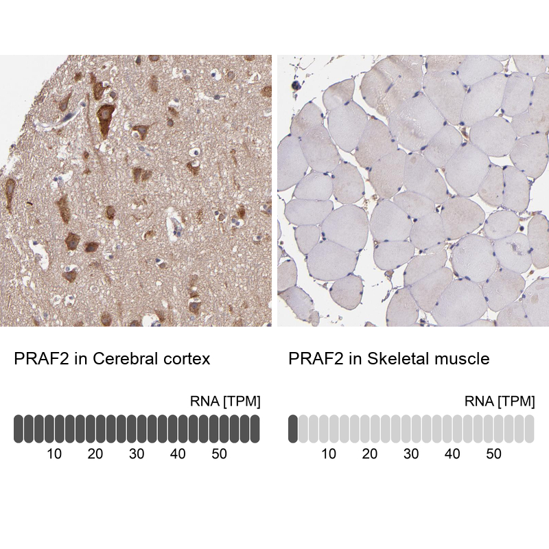
- Immunohistochemistry analysis in human cerebral cortex and skeletal muscle tissues using HPA002859 antibody. Corresponding PRAF2 RNA-seq data are presented for the same tissues.
- Sample type
- Human
- Protocol
- Protocol