 Explore
 Validate
 Learn
Learn Western blot
Antibody data
- Antibody Data
- Antigen structure
- References [3]
- Comments [0]
- Validations
- Western blot [1]
- Immunocytochemistry [1]
- Immunohistochemistry [2]
- Flow cytometry [6]
Submit
Validation data
Reference
Comment
Report error
- Product number
- MA1-82400 - Provider product page

- Provider
- Invitrogen Antibodies
- Product name
- CD20 Monoclonal Antibody (2H7)
- Antibody type
- Monoclonal
- Antigen
- Other
- Description
- For FACS analysis, use 10 µL of the suggested working dilution to label 1x10^6 cells in 100 µL. Mouse anti Human CD20 antibody, clone 2H7 recognizes the human CD20 cell surface antigen, a 33-37 kDa non-glycosylated phosphoprotein.
- Reactivity
- Human
- Host
- Mouse
- Isotype
- IgG
- Antibody clone number
- 2H7
- Vial size
- 200 µg
- Concentration
- 1 mg/mL
- Storage
- Store at 4°C short term. For long term storage, store at -20°C, avoiding freeze/thaw cycles.
Submitted references Mucosal Inducible NO Synthase-Producing IgA+ Plasma Cells in Helicobacter pylori-Infected Patients.
Effects of B Cell Depletion on Early Mycobacterium tuberculosis Infection in Cynomolgus Macaques.
Evidence for innate immune system activation in HIV type 1-infected elite controllers.
Neumann L, Mueller M, Moos V, Heller F, Meyer TF, Loddenkemper C, Bojarski C, Fehlings M, Doerner T, Allers K, Aebischer T, Ignatius R, Schneider T
Journal of immunology (Baltimore, Md. : 1950) 2016 Sep 1;197(5):1801-8
Journal of immunology (Baltimore, Md. : 1950) 2016 Sep 1;197(5):1801-8
Effects of B Cell Depletion on Early Mycobacterium tuberculosis Infection in Cynomolgus Macaques.
Phuah J, Wong EA, Gideon HP, Maiello P, Coleman MT, Hendricks MR, Ruden R, Cirrincione LR, Chan J, Lin PL, Flynn JL
Infection and immunity 2016 May;84(5):1301-1311
Infection and immunity 2016 May;84(5):1301-1311
Evidence for innate immune system activation in HIV type 1-infected elite controllers.
Krishnan S, Wilson EM, Sheikh V, Rupert A, Mendoza D, Yang J, Lempicki R, Migueles SA, Sereti I
The Journal of infectious diseases 2014 Mar;209(6):931-9
The Journal of infectious diseases 2014 Mar;209(6):931-9
No comments: Submit comment
Supportive validation
- Submitted by
- Invitrogen Antibodies (provider)
- Main image

- Experimental details
- Western blot was performed using Anti-CD20 Monoclonal Antibody (2H7) (Product # MA1-82400) and a 33 kDa band corresponding to CD20 was observed across relevant cell lines tested. Membrane enriched extracts (40 µg lysate) of Raji (Lane 1), Ramos (Lane 2), Jurkat (Lane 3), Hep G2 (Lane 4), Sp2/0-Ag14 (Lane 5), EL4 (Lane 6) were electrophoresed using NuPAGE™ 4-12% Bis-Tris Protein Gel (Product # NP0321BOX). Resolved proteins were then transferred onto a Nitrocellulose membrane (Product # IB23001) by iBlot® 2 Dry Blotting System (Product # IB21001). The blot was probed with the primary antibody (1:1000 dilution) and detected by chemiluminescence with Goat anti-Mouse IgG (H+L) Superclonal™ Recombinant Secondary Antibody, HRP (Product # A28177,1:4000 dilution) using the iBright FL 1000 (Product # A32752). Chemiluminescent detection was performed using SuperSignal™ West Dura Extended Duration Substrate (Product # 34076).

Supportive validation
- Submitted by
- Invitrogen Antibodies (provider)
- Main image

- Experimental details
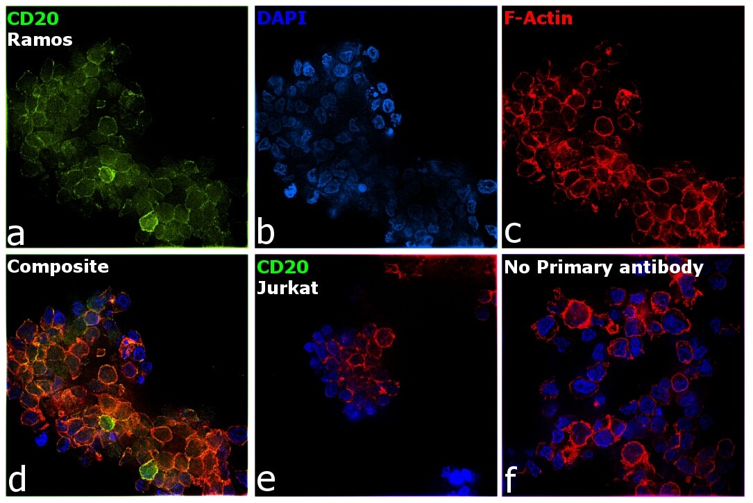
- Immunofluorescence analysis of CD20 was performed using 70% confluent log phase Ramos cells. The cells were fixed with 4% paraformaldehyde for 5 minutes, permeabilized with 0.1% Triton™ X-100 for 10 minutes, and blocked with 2% BSA for 45 minutes at room temperature. The cells were labeled with CD20 Monoclonal Antibody (2H7) (Product # MA1-82400) at 1:100 dilution in 0.1% BSA, incubated at 4 degree celsius overnight and then labeled with Donkey anti-Mouse IgG (H+L) Highly Cross-Adsorbed Secondary Antibody, Alexa Fluor Plus 488 (Product # A32766), (1:2000 dilution), for 45 minutes at room temperature (Panel a: Green). Nuclei (Panel b: Blue) were stained with ProLong™ Diamond Antifade Mountant with DAPI (Product # P36962). F-actin (Panel c: Red) was stained with Rhodamine Phalloidin (Product # R415, 1:300). Panel d represents the merged image showing plasma membrane and cytoplasm localization. Panel e represents negative cell line Jurkat. Panel f represents control cells with no primary antibody to assess background. The images were captured at 60X magnification.
Supportive validation
- Submitted by
- Invitrogen Antibodies (provider)
- Main image

- Experimental details
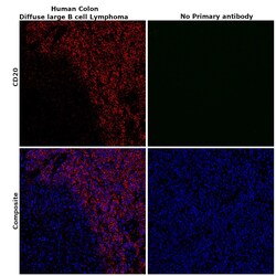
- Immunohistochemical analysis of CD20 was performed using formalin-fixed paraffin-embedded human colon (Diffuse large B cell lymphoma) tissue sections. To expose the target protein, heat-induced epitope retrieval was performed on de-paraffinized sections using eBioscience™ IHC Antigen Retrieval Solution - High pH (10X) (Product # 00-4956-58) diluted to 1X solution in water in a decloaking chamber at 110 degree Celsius for 15 minutes. Following antigen retrieval, the sections were blocked with 2% normal goat serum in 1X PBS for 45 minutes at room temperature and then probed with or without CD20 Monoclonal Antibody (2H7) (Product #MA1-82400) at 1:100 dilution in 0.1% normal goat serum overnight at 4 degree Celsius in a humidified chamber. Detection was performed using Goat anti-Mouse IgG (H+L) Highly Cross-Adsorbed Secondary Antibody, Alexa Fluor Plus 488 (Product # A32723) at a dilution of 1:2000 in 0.1% normal goat serum for 45 minutes at room temperature. ReadyProbes™ Tissue Autofluorescence Quenching Kit (Product # R37630) was used to quench autofluorescence from the tissues. Nuclei were stained with DAPI (Product # D1306) and the sections were mounted using ProLong™ Glass Antifade Mountant (Product # P36984). The images were captured on EVOS™ M7000 Imaging System (Product # AMF7000) at 20X magnification and externally deconvoluted.

- Submitted by
- Invitrogen Antibodies (provider)
- Main image
- Experimental details
- Immunohistochemical analysis of CD20 was performed using formalin-fixed paraffin-embedded human colon (Diffuse large B cell lymphoma) tissue sections. To expose the target protein, heat-induced epitope retrieval was performed on de-paraffinized sections using eBioscience™ IHC Antigen Retrieval Solution - High pH (10X) (Product # 00-4956-58) diluted to 1X solution in water in a decloaking chamber at 110 degree Celsius for 15 minutes. Following antigen retrieval, the sections were blocked with 2% normal goat serum in 1X PBS for 45 minutes at room temperature and then probed with or without CD20 Monoclonal Antibody (2H7) (Product #MA1-82400) at 1:100 dilution in 0.1% normal goat serum overnight at 4 degree Celsius in a humidified chamber. Detection was performed using Goat anti-Mouse IgG (H+L) Highly Cross-Adsorbed Secondary Antibody, Alexa Fluor Plus 488 (Product # A32723) at a dilution of 1:2000 in 0.1% normal goat serum for 45 minutes at room temperature. ReadyProbes™ Tissue Autofluorescence Quenching Kit (Product # R37630) was used to quench autofluorescence from the tissues. Nuclei were stained with DAPI (Product # D1306) and the sections were mounted using ProLong™ Glass Antifade Mountant (Product # P36984). The images were captured on EVOS™ M7000 Imaging System (Product # AMF7000) at 20X magnification and externally deconvoluted.

Supportive validation
- Submitted by
- Invitrogen Antibodies (provider)
- Main image

- Experimental details
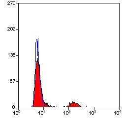
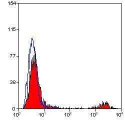
- Flow cytometric analysis of human peripheral blood lymphocytes using a CD20/MS4A1 monoclonal antibody (Product # MA1-82400)

- Submitted by
- Invitrogen Antibodies (provider)
- Main image

- Experimental details
- Flow cytometric analysis of human peripheral blood lymphocytes using a CD20/MS4A1 monoclonal antibody (Product # MA1-82400)

- Submitted by
- Invitrogen Antibodies (provider)
- Main image

- Experimental details
- Flow cytometric analysis of human peripheral blood lymphocytes using a CD20/MS4A1 monoclonal antibody (Product # MA1-82400)
- Submitted by
- Invitrogen Antibodies (provider)
- Main image

- Experimental details
- Flow cytometric analysis of human peripheral blood lymphocytes using a CD20/MS4A1 monoclonal antibody (Product # MA1-82400)

- Submitted by
- Invitrogen Antibodies (provider)
- Main image

- Experimental details
- Flow cytometric analysis of human peripheral blood lymphocytes using a CD20/MS4A1 monoclonal antibody (Product # MA1-82400)
- Submitted by
- Invitrogen Antibodies (provider)
- Main image

- Experimental details
- Flow cytometric analysis of human peripheral blood lymphocytes using a CD20/MS4A1 monoclonal antibody (Product # MA1-82400)
