Explore
 Validate
 Learn
Learn Western blot
Antibody data
- Antibody Data
- Antigen structure
- References [0]
- Comments [0]
- Validations
- Western blot [1]
- Flow cytometry [1]
Submit
Validation data
Reference
Comment
Report error
- Product number
- 10-4162-25 - Provider product page

- Provider
- ABEOMICS Inc.
- Product name
- Anti-CD20 Antibody
- Antibody type
- Monoclonal
- Description
- CD20 is clinically validated as an immunotherapy target for B-cell lymphomas and autoimmune diseases. CD20 consists of large, intracellular, amino- and carboxyterminal portions connected by 4 membrane-spanning domains. Its high expression on malignant B cells and its reported lack of shedding from the surfacemake CD20 an ideal target for antibody-mediated killing. Anti-CD20 antibodies are believed to mediate the therapeutic effect by activation of complement-dependent cytotoxicity (CDC) and largely by antibody-dependent cellular cytotoxicity exerted by recruitment of innate immune effector cells expressing the Fcgamma receptor IIIa.
- Reactivity
- Human
- Host
- Mouse
- Conjugate
- Unconjugated
- Antigen sequence
A partial length recombinant protei
n corresponding to extra cellular d
omain of CD20 was used as the immun
ogen for this antibody.- Isotype
- IgG
- Antibody clone number
- ABM46C7
- Vial size
- 100 µg
- Concentration
- 0.5 mg/ml
- Storage
- Store the antibody at 4°C, stable for 6 months. For long-term storage, store at -20°C. Avoid repeat freez thawing
No comments: Submit comment
Supportive validation
- Submitted by
- ABEOMICS Inc. (provider)
- Main image
- Experimental details
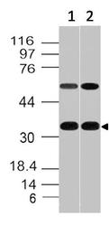
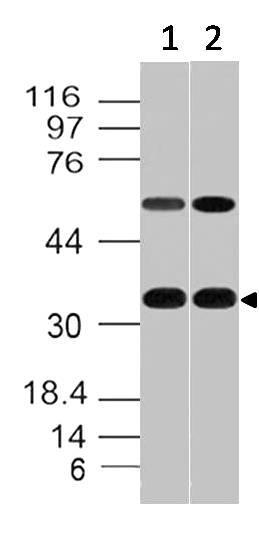
- Western blot analysis of CD20. Anti-CD20 antibody (Clone: ABM46C7) was tested at 2 µg/ml on Raji and Ramos lysates.
- Protocol
- Protocol
Supportive validation
- Submitted by
- ABEOMICS Inc. (provider)
- Main image

- Experimental details
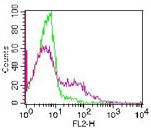
- Cell surface flow analysis of hCD20 on human PBMCs (Lymphocytes gated) using 0.5 µg/ 10^6 cells. Green represents isotype control (ABEOMICS); red represents anti-hCD20 antibody (10-4162). Goat anti-mouse PE conjugated secondary antibody (ABEOMICS) was used. (Cells were incubated with primary antibody for 45 min. then washed twice with FLOW Staining buffer (ABEOMICS) by centrifuging at 1100 rpm for 5 min, followed by 30 min incubation with conjugated secondary antibody. Data acquisition was done after washing twice with staining buffer).
- Protocol
- Protocol