 Explore
 Validate
 Learn
Learn Western blot
 Flow cytometry
Antibody data
- Antibody Data
- Antigen structure
- References [2]
- Comments [0]
- Validations
- Western blot [3]
- Immunocytochemistry [2]
- Immunohistochemistry [3]
Submit
Validation data
Reference
Comment
Report error
- Product number
- MA5-16334 - Provider product page

- Provider
- Invitrogen Antibodies
- Product name
- CD20 Monoclonal Antibody (SP32)
- Antibody type
- Monoclonal
- Antigen
- Synthetic peptide
- Description
- Heat-mediated antigen retrieval is recommended prior to staining, using a 10mM citrate buffer, pH 6.0, for 10 minutes followed by cooling at room temperature for 20 min. Following antigen retrieval, incubate samples with primary antibody for 30 min at room temperature. A suggested positive control is tonsil or B cell lymphoma.
- Reactivity
- Human, Mouse
- Host
- Rabbit
- Isotype
- IgG
- Antibody clone number
- SP32
- Vial size
- 500 µL
- Concentration
- Conc. not determined
- Storage
- -20° C, Avoid Freeze/Thaw Cycles
Submitted references Gut microbiota-specific IgA(+) B cells traffic to the CNS in active multiple sclerosis.
Characterization of post transplantation lymphoma in feline renal transplant recipients.
Pröbstel AK, Zhou X, Baumann R, Wischnewski S, Kutza M, Rojas OL, Sellrie K, Bischof A, Kim K, Ramesh A, Dandekar R, Greenfield AL, Schubert RD, Bisanz JE, Vistnes S, Khaleghi K, Landefeld J, Kirkish G, Liesche-Starnecker F, Ramaglia V, Singh S, Tran EB, Barba P, Zorn K, Oechtering J, Forsberg K, Shiow LR, Henry RG, Graves J, Cree BAC, Hauser SL, Kuhle J, Gelfand JM, Andersen PM, Schlegel J, Turnbaugh PJ, Seeberger PH, Gommerman JL, Wilson MR, Schirmer L, Baranzini SE
Science immunology 2020 Nov 20;5(53)
Science immunology 2020 Nov 20;5(53)
Characterization of post transplantation lymphoma in feline renal transplant recipients.
Durham AC, Mariano AD, Holmes ES, Aronson L
Journal of comparative pathology 2014 Feb-Apr;150(2-3):162-8
Journal of comparative pathology 2014 Feb-Apr;150(2-3):162-8
No comments: Submit comment
Supportive validation
- Submitted by
- Invitrogen Antibodies (provider)
- Main image

- Experimental details
- Western blot analysis of Ramos Cells using anti-CD20 Monoclonal Antibody (Product # MA5-16334). The recommened dilution for this antibody in western blot applications is 1:100.

- Submitted by
- Invitrogen Antibodies (provider)
- Main image

- Experimental details
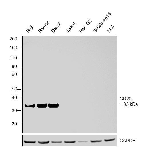
- Western blot was performed using Anti-CD20 Monoclonal Antibody (SP32) (Product # MA1-39416) and a 33 kDa band corresponding to CD20 was observed across relevant cell lines tested. Membrane enriched extracts (40 µg lysate) of Raji (Lane 1), Ramos (Lane 2), Daudi (Lane 3), Jurkat (Lane 4), Hep G2 (Lane 5), Sp2/0-Ag14 (Lane 6), EL4 (Lane 7) were electrophoresed using NuPAGE™ 4-12% Bis-Tris Protein Gel (Product # NP0321BOX). Resolved proteins were then transferred onto a Nitrocellulose membrane (Product # IB23001) by iBlot® 2 Dry Blotting System (Product # IB21001). The blot was probed with the primary antibody (1:500 dilution) and detected by chemiluminescence with Goat anti-Rabbit IgG (H+L) Superclonal™ Recombinant Secondary Antibody, HRP (Product # A27036,1:4000 dilution) using the iBright FL 1000 (Product # A32752). Chemiluminescent detection was performed using Novex® ECL Chemiluminescent Substrate Reagent Kit (Product # WP20005).
- Submitted by
- Invitrogen Antibodies (provider)
- Main image

- Experimental details
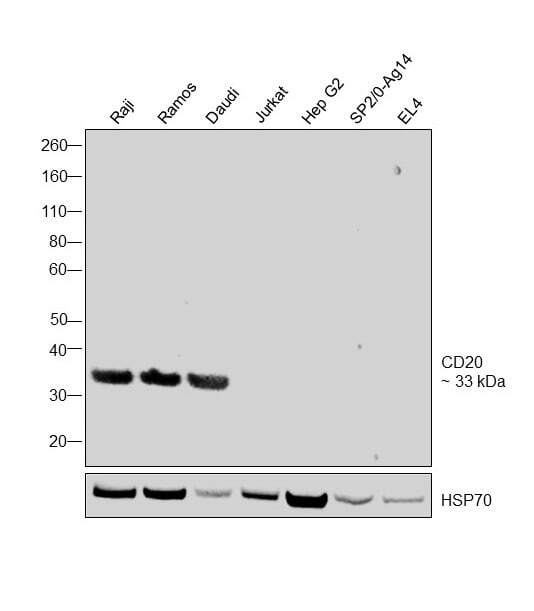
- Western blot was performed using Anti-CD20 Monoclonal Antibody (SP32) (Product # MA5-16334) and a 33 kDa band corresponding to CD20 was observed in Raji, Ramos, Daudi and undetected in Jurkat, HepG2 and mouse cell lines tested. Membrane enriched cell lysate (40 µg lysate) of Raji (Lane 1), Ramos (Lane 2), Daudi (Lane 3), Jurkat (Lane 4), Hep G2 (Lane 5), Sp2/0-Ag14 (Lane 6), EL4 (Lane 7) were electrophoresed using NuPAGE™ 4-12% Bis-Tris Protein Gel (Product # NP0321BOX). Resolved proteins were then transferred onto a Nitrocellulose membrane (Product # IB23001) by iBlot® 2 Dry Blotting System (Product # IB21001). The blot was probed with the primary antibody (1:500 dilution) and detected by chemiluminescence with Goat anti-Rabbit IgG (H+L) Superclonal™ Recombinant Secondary Antibody, HRP (Product # A27036, 1:4000 dilution) using the iBright FL 1000 (Product # A32752). Chemiluminescent detection was performed using Novex® ECL Chemiluminescent Substrate Reagent Kit (Product # WP20005).
Supportive validation
- Submitted by
- Invitrogen Antibodies (provider)
- Main image
- Experimental details
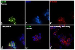
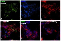
- Immunofluorescence analysis of CD20 was performed using 70% confluent log phase Ramos cells. The cells were fixed with 4% paraformaldehyde for 5 minutes, permeabilized with 0.1% Triton™ X-100 for 10 minutes, and blocked with 2% BSA for 45 minutes at room temperature. The cells were labeled with CD20 Monoclonal Antibody (SP32) (Product # MA1-39416) at 1:100 dilution in 0.1% BSA, incubated at 4 degree celsius overnight and then labeled with Donkey anti-Rabbit IgG (H+L) Highly Cross-Adsorbed Secondary Antibody, Alexa Fluor Plus 488 (Product # A32790), (1:2000 dilution), for 45 minutes at room temperature (Panel a: Green). Nuclei (Panel b: Blue) were stained with ProLong™ Diamond Antifade Mountant with DAPI (Product # P36962). F-actin (Panel c: Red) was stained with Rhodamine Phalloidin (Product # R415, 1:300). Panel d represents the merged image showing plasma membrane localization. Panel e represents negative cell line Jurkat. Panel f represents control cells with no primary antibody to assess background. The images were captured at 60X magnification.
- Submitted by
- Invitrogen Antibodies (provider)
- Main image
- Experimental details
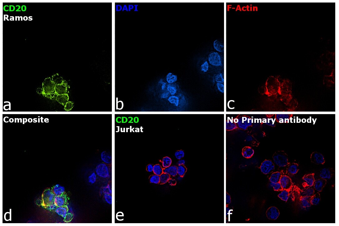
- Immunofluorescence analysis of CD20 was performed using 70 % confluent log phase Ramos cells. The cells were fixed with 4% paraformaldehyde for 5 minutes, permeabilized with 0.1% Triton™ X-100 for 10 minutes, and blocked with 2% BSA for 45 minutes at room temperature. The cells were labeled with CD20 Monoclonal Antibody (SP32) (Product # MA5-16334) at 1:100 dilution in 0.1% BSA, incubated at 4 degree celsius overnight and then labeled with Donkey anti-Rabbit IgG (H+L) Highly Cross-Adsorbed Secondary Antibody, Alexa Fluor Plus 555 (Product # A32794), (1:2000 dilution), for 45 minutes at room temperature (Panel a: Green). Nuclei (Panel b: Blue) were stained with ProLong™ Diamond Antifade Mountant with DAPI (Product # P36962). F-actin (Panel c: Red) was stained with Rhodamine Phalloidin (Product # R415, 1:300). Panel d represents the merged image showing plasma membrane localization. Panel e represents negative cell line Jurkat. Panel f represents control cells with no primary antibody to assess background. The images were captured at 60X magnification.

Supportive validation
- Submitted by
- Invitrogen Antibodies (provider)
- Main image

- Experimental details
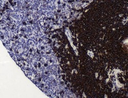
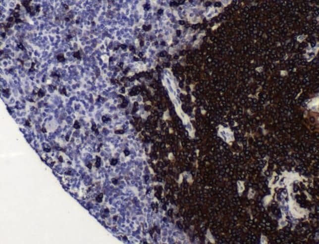
- Immunohistochemistry analysis was performed on elephant lymph node tissues. To expose target proteins, antigen retrieval was performed by pressure cooker for 30 minutes in DIVA decloaker (pH 6.0). Following antigen retrieval, endogenous peroxidases were blocked with 3% hydrogen peroxide for 10 min at room temperature. Tissue slides were washed with TBS-T, and then blocked in goat-serum for 30 min at room temperature. Tissues were probed with CD20 monoclonal antibody (MA5-16334) at 1:100 for 1 hour at 4°C in a humidified chamber. Tissues were washed extensively and detection was performed using a biotinylated anti-rabbit secondary antibody and avidin-conjugated to horseradish peroxidase, then followed by colorimetric detection using DAB. Tissues were counterstained with hematoxylin and dehydrated with ethanol and xylene to prep for mounting. Images were taken on an Olympus BX-43 microscope.

- Submitted by
- Invitrogen Antibodies (provider)
- Main image
- Experimental details
- Immunohistochemical analysis of CD20 using anti-CD20 Monoclonal Antibody (Product # MA5-16334) in B Cell Lymphoma Cancer Tissue. The recommened dilution for this antibody in immunohistochemistry applications is 1:100.
- Submitted by
- Invitrogen Antibodies (provider)
- Main image

- Experimental details
- Immunohistochemical analysis of CD20 using a monoclonal antibody (Product # MA1-39416).
