Explore
 Validate
 Learn
Learn Western blot
Antibody data
- Antibody Data
- Antigen structure
- References [67]
- Comments [0]
- Validations
- Western blot [2]
- Immunocytochemistry [2]
- Immunohistochemistry [8]
- Flow cytometry [2]
- Other assay [20]
Submit
Validation data
Reference
Comment
Report error
- Product number
- PA5-16701 - Provider product page

- Provider
- Invitrogen Antibodies
- Product name
- CD20 Polyclonal Antibody
- Antibody type
- Polyclonal
- Antigen
- Synthetic peptide
- Description
- Upon delivery aliquot and store at -20ºC, avoiding freeze/thaw cycles. For IHC staining, antigen retrieval is not essential but may optimize staining. Recommended positive controls: BJAB WCL and tonsil.
- Reactivity
- Human, Mouse, Canine, Feline
- Host
- Rabbit
- Isotype
- IgG
- Vial size
- 500 µL
- Concentration
- 0.077 mg/mL
- Storage
- -20°C, Avoid Freeze/Thaw Cycles
Submitted references Inflammatory Related Reactions in Humans and in Canine Breast Cancers, A Spontaneous Animal Model of Disease.
Rapamycin-filgrastim combination therapy ameliorates portal hypertension-induced splenomegaly: Role of β actin and S100A9 proteins modulation.
Distinct histopathological phenotypes of severe alcoholic hepatitis suggest different mechanisms driving liver injury and failure.
A rapid multiple immunofluorescence method for the simultaneous detection of CD20 and CD3 in canine and feline cytological samples.
Low-dose Ad26.COV2.S protection against SARS-CoV-2 challenge in rhesus macaques.
Gene dosage effects of poly(A) track-engineered hypomorphs.
Pharmacological Inhibition of MALT1 Protease Leads to a Progressive IPEX-Like Pathology.
Efficacy of Local Minocycline Agents in Treating Peri-Implantitis: An Experimental In Vivo Study in Beagle Dogs.
Local Lung Immune Response to Mycobacterium bovis Challenge after BCG and M. bovis Heat-Inactivated Vaccination in European Badger (Meles meles).
Multifocal cutaneous non-epitheliotropic B-cell lymphoma in a cat.
Immunohistochemical Assessment of Immune Response in the Dermis of Sarcoptes scabiei-Infested Wild Carnivores (Wolf and Fox) and Ruminants (Chamois and Red Deer).
Morphological and Immunohistochemical Examination of Lymphoproliferative Lesions Caused by Marek's Disease Virus in Breeder Chickens.
Neuroinflammation is associated with infiltration of T cells in Lewy body disease and α-synuclein transgenic models.
Locally Applied Slow-Release of Minocycline Microspheres in the Treatment of Peri-Implant Mucositis: An Experimental In Vivo Study.
Lymphocyte Populations in the Adventitial Layer of Hydatid Cysts in Cattle: Relationship With Cyst Fertility Status and Fasciola Hepatica Co-Infection.
Phenotypic Characterization of Encephalitis and Immune Response in the Brains of Lambs Experimentally Infected with Spanish Goat Encephalitis Virus.
Prostaglandin E(2)-Induced Immune Exhaustion and Enhancement of Antiviral Effects by Anti-PD-L1 Antibody Combined with COX-2 Inhibitor in Bovine Leukemia Virus Infection.
A truncating mutation in the autophagy gene UVRAG drives inflammation and tumorigenesis in mice.
Toxicology Study of Intra-Cisterna Magna Adeno-Associated Virus 9 Expressing Human Alpha-L-Iduronidase in Rhesus Macaques.
Toxicology Study of Intra-Cisterna Magna Adeno-Associated Virus 9 Expressing Iduronate-2-Sulfatase in Rhesus Macaques.
Characterization of a Vesivirus Associated with an Outbreak of Acute Hemorrhagic Gastroenteritis in Domestic Dogs.
A small, portable RNA device for the control of exon skipping in mammalian cells.
Severe Toxicity in Nonhuman Primates and Piglets Following High-Dose Intravenous Administration of an Adeno-Associated Virus Vector Expressing Human SMN.
B cell-derived IL-4 acts on podocytes to induce proteinuria and foot process effacement.
Dynamic Autologous Reendothelialization of Small-Caliber Arterial Extracellular Matrix: A Preclinical Large Animal Study.
Clinical and histopathological evaluation of 16 dogs with T-zone lymphoma.
Evaluation of CD25-positive cells in relation to the subtypes and prognoses in various lymphoid tumours in dogs.
Therapeutic Efficacy of Fresh, Autologous Mesenchymal Stem Cells for Severe Refractory Gingivostomatitis in Cats.
ZEB2 drives immature T-cell lymphoblastic leukaemia development via enhanced tumour-initiating potential and IL-7 receptor signalling.
MMP-9 expression is increased in B lymphocytes during multiple sclerosis exacerbation and is regulated by microRNA-320a.
Loss of PTPN12 Stimulates Progression of ErbB2-Dependent Breast Cancer by Enhancing Cell Survival, Migration, and Epithelial-to-Mesenchymal Transition.
Canine indolent and aggressive lymphoma: clinical spectrum with histologic correlation.
Heterogeneous pathological outcomes after experimental pH1N1 influenza infection in ferrets correlate with viral replication and host immune responses in the lung.
Pathology of fatal lineage 1 and 2 West Nile virus infections in horses in South Africa.
Regulation of the development of asthmatic inflammation by in situ CD4(+)Foxp3 (+) T cells in a mouse model of late allergic asthma.
Intraocular and periocular lymphoma in dogs and cats: a retrospective review of 21 cases (2001-2012).
Immunohistochemical expression of cyclooxygenase-2 in normal, hyperplastic and neoplastic canine lymphoid tissues.
Spinal meningeal oligodendrogliomatosis in two boxer dogs.
Assessment of bone marrow infiltration diagnosed by flow cytometry in canine large B cell lymphoma: prognostic significance and proposal of a cut-off value.
The expression ratio of miR-17-5p and miR-155 correlates with grading in canine splenic lymphoma.
Digital dermatitis in cattle is associated with an excessive innate immune response triggered by the keratinocytes.
Granule exocytosis of granulysin and granzyme B as a potential key mechanism in vaccine-induced immunity in cattle against the nematode Ostertagia ostertagi.
Equine multisystemic eosinophilic epitheliotropic disease: a case report and review of literature.
B-cell lymphoma with plasmacytoid differentiation, atypical cytoplasmic inclusions, and secondary leukemia in a dog.
The dog as a possible animal model for human non-Hodgkin lymphoma: a review.
Clinical characteristics and outcome in dogs with splenic marginal zone lymphoma.
Canine inflamed nonepitheliotropic cutaneous T-cell lymphoma: a diagnostic conundrum.
Congenital ascites due to hepatoblastoma with extensive peritoneal implantation metastases in a premature equine fetus.
Safety and immunomodulatory effects of allogeneic canine adipose-derived mesenchymal stromal cells transplanted into the region of the lacrimal gland, the gland of the third eyelid and the knee joint.
BLT-humanized C57BL/6 Rag2-/-γc-/-CD47-/- mice are resistant to GVHD and develop B- and T-cell immunity to HIV infection.
Development and characterization of 5 canine B-cell lymphoma cell lines.
A retrospective study of the clinical, histological, and immunohistochemical manifestations of 5 dogs originally diagnosed histologically as necrotizing scleritis.
Vaccination reduces macrophage infiltration in bronchus-associated lymphoid tissue in pigs infected with a highly virulent Mycoplasma hyopneumoniae strain.
CD20+ tumor-infiltrating lymphocytes have an atypical CD27- memory phenotype and together with CD8+ T cells promote favorable prognosis in ovarian cancer.
Diagnostic yield and adverse effects of MRI-guided free-hand brain biopsies through a mini-burr hole in dogs with encephalitis.
Intramuscular inoculation of cattle with Sarcocystis antigen results in focal eosinophilic myositis.
Intratumoral Immune Responses Can Distinguish New Primary and True Recurrence Types of Ipsilateral Breast Tumor Recurrences (IBTR).
Tumor-infiltrating lymphocytes predict response to anthracycline-based chemotherapy in estrogen receptor-negative breast cancer.
Innate immune activation potentiates alloimmune lung disease independent of chemokine (C-X-C motif) receptor 3.
Use of tissue microarray and automated quantitative analysis for screening and validation of potential biomarkers in mantle cell lymphoma.
Hemophagocytic histiocytic sarcoma in a Japanese black cow.
Tumor-infiltrating T cells correlate with NY-ESO-1-specific autoantibodies in ovarian cancer.
Feline pulmonary Langerhans cell histiocytosis with multiorgan involvement.
Allogeneic lymphocytes persist and traffic in feral MHC-matched mauritian cynomolgus macaques.
B-cell intestinal lymphoma with Mott cell differentiation in a 1-year-old miniature Dachshund.
Proliferation of immature plasma cells in pouchitis mucosa in patients with ulcerative colitis.
A novel canine lymphoma cell line: a translational and comparative model for lymphoma research.
Tricarico D, Convertino AS, Mehmeti I, Ranieri G, Leonetti F, Laface C, Zizzo N
Frontiers in pharmacology 2022;13:752098
Frontiers in pharmacology 2022;13:752098
Rapamycin-filgrastim combination therapy ameliorates portal hypertension-induced splenomegaly: Role of β actin and S100A9 proteins modulation.
Abdelrahman SA, Abdelfatah MM, Keshta AT
Iranian journal of basic medical sciences 2022 Jun;25(6):732-744
Iranian journal of basic medical sciences 2022 Jun;25(6):732-744
Distinct histopathological phenotypes of severe alcoholic hepatitis suggest different mechanisms driving liver injury and failure.
Ma J, Guillot A, Yang Z, Mackowiak B, Hwang S, Park O, Peiffer BJ, Ahmadi AR, Melo L, Kusumanchi P, Huda N, Saxena R, He Y, Guan Y, Feng D, Sancho-Bru P, Zang M, Cameron AM, Bataller R, Tacke F, Sun Z, Liangpunsakul S, Gao B
The Journal of clinical investigation 2022 Jul 15;132(14)
The Journal of clinical investigation 2022 Jul 15;132(14)
A rapid multiple immunofluorescence method for the simultaneous detection of CD20 and CD3 in canine and feline cytological samples.
Matsumoto F, Shima-Sawa M, Furusawa Y, Yamato O, Yabuki A
The Journal of veterinary medical science 2021 May 17;83(5):832-836
The Journal of veterinary medical science 2021 May 17;83(5):832-836
Low-dose Ad26.COV2.S protection against SARS-CoV-2 challenge in rhesus macaques.
He X, Chandrashekar A, Zahn R, Wegmann F, Yu J, Mercado NB, McMahan K, Martinot AJ, Piedra-Mora C, Beecy S, Ducat S, Chamanza R, Huber SR, van Heerden M, van der Fits L, Borducchi EN, Lifton M, Liu J, Nampanya F, Patel S, Peter L, Tostanoski LH, Pessaint L, Van Ry A, Finneyfrock B, Velasco J, Teow E, Brown R, Cook A, Andersen H, Lewis MG, Schuitemaker H, Barouch DH
Cell 2021 Jun 24;184(13):3467-3473.e11
Cell 2021 Jun 24;184(13):3467-3473.e11
Gene dosage effects of poly(A) track-engineered hypomorphs.
Powell G, Pavlovic Djuranovic S, Djuranovic S
Molecular therapy. Nucleic acids 2021 Dec 3;26:865-878
Molecular therapy. Nucleic acids 2021 Dec 3;26:865-878
Pharmacological Inhibition of MALT1 Protease Leads to a Progressive IPEX-Like Pathology.
Martin K, Junker U, Tritto E, Sutter E, Rubic-Schneider T, Morgan H, Niwa S, Li J, Schlapbach A, Walker D, Bigaud M, Beerli C, Littlewood-Evans A, Rudolph B, Laisney M, Ledieu D, Beltz K, Quancard J, Bornancin F, Zamurovic Ribrioux N, Calzascia T
Frontiers in immunology 2020;11:745
Frontiers in immunology 2020;11:745
Efficacy of Local Minocycline Agents in Treating Peri-Implantitis: An Experimental In Vivo Study in Beagle Dogs.
Yoon SW, Kim MJ, Paeng KW, Yu KA, Lee CK, Song YW, Cha JK, Jung UW
Pharmaceutics 2020 Oct 23;12(11)
Pharmaceutics 2020 Oct 23;12(11)
Local Lung Immune Response to Mycobacterium bovis Challenge after BCG and M. bovis Heat-Inactivated Vaccination in European Badger (Meles meles).
Blanco Vázquez C, Prieto M, Barral M, Juste RA, Lesellier S, Salguero FJ, Davé D, Martínez IZ, de Garnica García MG, Casais R, Balseiro A
Pathogens (Basel, Switzerland) 2020 Jun 9;9(6)
Pathogens (Basel, Switzerland) 2020 Jun 9;9(6)
Multifocal cutaneous non-epitheliotropic B-cell lymphoma in a cat.
Quintavalla F, Di Lecce R, Carlini D, Zanfabro M, Cantoni AM
JFMS open reports 2020 Jul-Dec;6(2):2055116920972077
JFMS open reports 2020 Jul-Dec;6(2):2055116920972077
Immunohistochemical Assessment of Immune Response in the Dermis of Sarcoptes scabiei-Infested Wild Carnivores (Wolf and Fox) and Ruminants (Chamois and Red Deer).
Martínez IZ, Oleaga Á, Sojo I, García-Iglesias MJ, Pérez-Martínez C, García Marín JF, Balseiro A
Animals : an open access journal from MDPI 2020 Jul 6;10(7)
Animals : an open access journal from MDPI 2020 Jul 6;10(7)
Morphological and Immunohistochemical Examination of Lymphoproliferative Lesions Caused by Marek's Disease Virus in Breeder Chickens.
Stamilla A, Messina A, Condorelli L, Licitra F, Antoci F, Lanza M, Loria GR, Cascone G, Puleio R
Animals : an open access journal from MDPI 2020 Jul 27;10(8)
Animals : an open access journal from MDPI 2020 Jul 27;10(8)
Neuroinflammation is associated with infiltration of T cells in Lewy body disease and α-synuclein transgenic models.
Iba M, Kim C, Sallin M, Kwon S, Verma A, Overk C, Rissman RA, Sen R, Sen JM, Masliah E
Journal of neuroinflammation 2020 Jul 17;17(1):214
Journal of neuroinflammation 2020 Jul 17;17(1):214
Locally Applied Slow-Release of Minocycline Microspheres in the Treatment of Peri-Implant Mucositis: An Experimental In Vivo Study.
Yoon SW, Kim MJ, Paeng KW, Yu KA, Lee CK, Song YW, Cha JK, Sanz M, Jung UW
Pharmaceutics 2020 Jul 16;12(7)
Pharmaceutics 2020 Jul 16;12(7)
Lymphocyte Populations in the Adventitial Layer of Hydatid Cysts in Cattle: Relationship With Cyst Fertility Status and Fasciola Hepatica Co-Infection.
Jiménez M, Stoore C, Hidalgo C, Corrêa F, Hernández M, Benavides J, Ferreras MC, Sáenz L, Paredes R
Veterinary pathology 2020 Jan;57(1):108-114
Veterinary pathology 2020 Jan;57(1):108-114
Phenotypic Characterization of Encephalitis and Immune Response in the Brains of Lambs Experimentally Infected with Spanish Goat Encephalitis Virus.
Martínez IZ, Pérez-Martínez C, Salinas LM, Juste RA, Marín JFG, Balseiro A
Animals : an open access journal from MDPI 2020 Aug 7;10(8)
Animals : an open access journal from MDPI 2020 Aug 7;10(8)
Prostaglandin E(2)-Induced Immune Exhaustion and Enhancement of Antiviral Effects by Anti-PD-L1 Antibody Combined with COX-2 Inhibitor in Bovine Leukemia Virus Infection.
Sajiki Y, Konnai S, Okagawa T, Nishimori A, Maekawa N, Goto S, Watari K, Minato E, Kobayashi A, Kohara J, Yamada S, Kaneko MK, Kato Y, Takahashi H, Terasaki N, Takeda A, Yamamoto K, Toda M, Suzuki Y, Murata S, Ohashi K
Journal of immunology (Baltimore, Md. : 1950) 2019 Sep 1;203(5):1313-1324
Journal of immunology (Baltimore, Md. : 1950) 2019 Sep 1;203(5):1313-1324
A truncating mutation in the autophagy gene UVRAG drives inflammation and tumorigenesis in mice.
Quach C, Song Y, Guo H, Li S, Maazi H, Fung M, Sands N, O'Connell D, Restrepo-Vassalli S, Chai B, Nemecio D, Punj V, Akbari O, Idos GE, Mumenthaler SM, Wu N, Martin SE, Hagiya A, Hicks J, Cui H, Liang C
Nature communications 2019 Dec 12;10(1):5681
Nature communications 2019 Dec 12;10(1):5681
Toxicology Study of Intra-Cisterna Magna Adeno-Associated Virus 9 Expressing Human Alpha-L-Iduronidase in Rhesus Macaques.
Hordeaux J, Hinderer C, Goode T, Katz N, Buza EL, Bell P, Calcedo R, Richman LK, Wilson JM
Molecular therapy. Methods & clinical development 2018 Sep 21;10:79-88
Molecular therapy. Methods & clinical development 2018 Sep 21;10:79-88
Toxicology Study of Intra-Cisterna Magna Adeno-Associated Virus 9 Expressing Iduronate-2-Sulfatase in Rhesus Macaques.
Hordeaux J, Hinderer C, Goode T, Buza EL, Bell P, Calcedo R, Richman LK, Wilson JM
Molecular therapy. Methods & clinical development 2018 Sep 21;10:68-78
Molecular therapy. Methods & clinical development 2018 Sep 21;10:68-78
Characterization of a Vesivirus Associated with an Outbreak of Acute Hemorrhagic Gastroenteritis in Domestic Dogs.
Renshaw RW, Griffing J, Weisman J, Crofton LM, Laverack MA, Poston RP, Duhamel GE, Dubovi EJ
Journal of clinical microbiology 2018 May;56(5)
Journal of clinical microbiology 2018 May;56(5)
A small, portable RNA device for the control of exon skipping in mammalian cells.
Vogel M, Weigand JE, Kluge B, Grez M, Suess B
Nucleic acids research 2018 May 4;46(8):e48
Nucleic acids research 2018 May 4;46(8):e48
Severe Toxicity in Nonhuman Primates and Piglets Following High-Dose Intravenous Administration of an Adeno-Associated Virus Vector Expressing Human SMN.
Hinderer C, Katz N, Buza EL, Dyer C, Goode T, Bell P, Richman LK, Wilson JM
Human gene therapy 2018 Mar;29(3):285-298
Human gene therapy 2018 Mar;29(3):285-298
B cell-derived IL-4 acts on podocytes to induce proteinuria and foot process effacement.
Kim AH, Chung JJ, Akilesh S, Koziell A, Jain S, Hodgin JB, Miller MJ, Stappenbeck TS, Miner JH, Shaw AS
JCI insight 2017 Nov 2;2(21)
JCI insight 2017 Nov 2;2(21)
Dynamic Autologous Reendothelialization of Small-Caliber Arterial Extracellular Matrix: A Preclinical Large Animal Study.
Dahan N, Sarig U, Bronshtein T, Baruch L, Karram T, Hoffman A, Machluf M
Tissue engineering. Part A 2017 Jan;23(1-2):69-79
Tissue engineering. Part A 2017 Jan;23(1-2):69-79
Clinical and histopathological evaluation of 16 dogs with T-zone lymphoma.
Mizutani N, Goto-Koshino Y, Takahashi M, Uchida K, Tsujimoto H
The Journal of veterinary medical science 2016 Sep 1;78(8):1237-44
The Journal of veterinary medical science 2016 Sep 1;78(8):1237-44
Evaluation of CD25-positive cells in relation to the subtypes and prognoses in various lymphoid tumours in dogs.
Mizutani N, Goto-Koshino Y, Tsuboi M, Kagawa Y, Ohno K, Uchida K, Tsujimoto H
Veterinary immunology and immunopathology 2016 May;173:39-43
Veterinary immunology and immunopathology 2016 May;173:39-43
Therapeutic Efficacy of Fresh, Autologous Mesenchymal Stem Cells for Severe Refractory Gingivostomatitis in Cats.
Arzi B, Mills-Ko E, Verstraete FJ, Kol A, Walker NJ, Badgley MR, Fazel N, Murphy WJ, Vapniarsky N, Borjesson DL
Stem cells translational medicine 2016 Jan;5(1):75-86
Stem cells translational medicine 2016 Jan;5(1):75-86
ZEB2 drives immature T-cell lymphoblastic leukaemia development via enhanced tumour-initiating potential and IL-7 receptor signalling.
Goossens S, Radaelli E, Blanchet O, Durinck K, Van der Meulen J, Peirs S, Taghon T, Tremblay CS, Costa M, Farhang Ghahremani M, De Medts J, Bartunkova S, Haigh K, Schwab C, Farla N, Pieters T, Matthijssens F, Van Roy N, Best JA, Deswarte K, Bogaert P, Carmichael C, Rickard A, Suryani S, Bracken LS, Alserihi R, Canté-Barrett K, Haenebalcke L, Clappier E, Rondou P, Slowicka K, Huylebroeck D, Goldrath AW, Janzen V, McCormack MP, Lock RB, Curtis DJ, Harrison C, Berx G, Speleman F, Meijerink JP, Soulier J, Van Vlierberghe P, Haigh JJ
Nature communications 2015 Jan 7;6:5794
Nature communications 2015 Jan 7;6:5794
MMP-9 expression is increased in B lymphocytes during multiple sclerosis exacerbation and is regulated by microRNA-320a.
Aung LL, Mouradian MM, Dhib-Jalbut S, Balashov KE
Journal of neuroimmunology 2015 Jan 15;278:185-9
Journal of neuroimmunology 2015 Jan 15;278:185-9
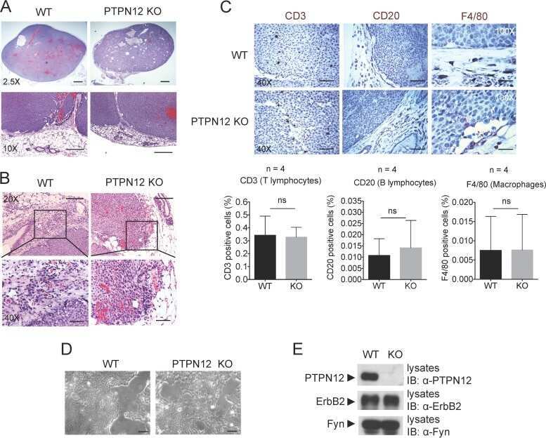
Loss of PTPN12 Stimulates Progression of ErbB2-Dependent Breast Cancer by Enhancing Cell Survival, Migration, and Epithelial-to-Mesenchymal Transition.
Li J, Davidson D, Martins Souza C, Zhong MC, Wu N, Park M, Muller WJ, Veillette A
Molecular and cellular biology 2015 Dec;35(23):4069-82
Molecular and cellular biology 2015 Dec;35(23):4069-82
Canine indolent and aggressive lymphoma: clinical spectrum with histologic correlation.
Aresu L, Martini V, Rossi F, Vignoli M, Sampaolo M, Aricò A, Laganga P, Pierini A, Frayssinet P, Mantovani R, Marconato L
Veterinary and comparative oncology 2015 Dec;13(4):348-62
Veterinary and comparative oncology 2015 Dec;13(4):348-62
Heterogeneous pathological outcomes after experimental pH1N1 influenza infection in ferrets correlate with viral replication and host immune responses in the lung.
Vidaña B, Martínez J, Martínez-Orellana P, García Migura L, Montoya M, Martorell J, Majó N
Veterinary research 2014;45(1):85
Veterinary research 2014;45(1):85
Pathology of fatal lineage 1 and 2 West Nile virus infections in horses in South Africa.
Williams JH, van Niekerk S, Human S, van Wilpe E, Venter M
Journal of the South African Veterinary Association 2014 Sep 1;85(1):1105
Journal of the South African Veterinary Association 2014 Sep 1;85(1):1105
Regulation of the development of asthmatic inflammation by in situ CD4(+)Foxp3 (+) T cells in a mouse model of late allergic asthma.
Nakashima T, Hayashi T, Mizuno T
Inflammation 2014 Oct;37(5):1642-53
Inflammation 2014 Oct;37(5):1642-53
Intraocular and periocular lymphoma in dogs and cats: a retrospective review of 21 cases (2001-2012).
Ota-Kuroki J, Ragsdale JM, Bawa B, Wakamatsu N, Kuroki K
Veterinary ophthalmology 2014 Nov;17(6):389-96
Veterinary ophthalmology 2014 Nov;17(6):389-96
Immunohistochemical expression of cyclooxygenase-2 in normal, hyperplastic and neoplastic canine lymphoid tissues.
Asproni P, Vignoli M, Cancedda S, Millanta F, Terragni R, Poli A
Journal of comparative pathology 2014 Jul;151(1):35-41
Journal of comparative pathology 2014 Jul;151(1):35-41
Spinal meningeal oligodendrogliomatosis in two boxer dogs.
Kovi RC, Wünschmann A, Armién AG, Hall K, Carlson T, Shivers J, Oglesbee MJ
Veterinary pathology 2013 Sep;50(5):761-4
Veterinary pathology 2013 Sep;50(5):761-4
Assessment of bone marrow infiltration diagnosed by flow cytometry in canine large B cell lymphoma: prognostic significance and proposal of a cut-off value.
Marconato L, Martini V, Aresu L, Sampaolo M, Valentini F, Rinaldi V, Comazzi S
Veterinary journal (London, England : 1997) 2013 Sep;197(3):776-81
Veterinary journal (London, England : 1997) 2013 Sep;197(3):776-81
The expression ratio of miR-17-5p and miR-155 correlates with grading in canine splenic lymphoma.
Albonico F, Mortarino M, Avallone G, Gioia G, Comazzi S, Roccabianca P
Veterinary immunology and immunopathology 2013 Sep 1;155(1-2):117-23
Veterinary immunology and immunopathology 2013 Sep 1;155(1-2):117-23
Digital dermatitis in cattle is associated with an excessive innate immune response triggered by the keratinocytes.
Refaai W, Ducatelle R, Geldhof P, Mihi B, El-shair M, Opsomer G
BMC veterinary research 2013 Oct 3;9:193
BMC veterinary research 2013 Oct 3;9:193
Granule exocytosis of granulysin and granzyme B as a potential key mechanism in vaccine-induced immunity in cattle against the nematode Ostertagia ostertagi.
Van Meulder F, Van Coppernolle S, Borloo J, Rinaldi M, Li RW, Chiers K, Van den Broeck W, Vercruysse J, Claerebout E, Geldhof P
Infection and immunity 2013 May;81(5):1798-809
Infection and immunity 2013 May;81(5):1798-809
Equine multisystemic eosinophilic epitheliotropic disease: a case report and review of literature.
Bosseler L, Verryken K, Bauwens C, de Vries C, Deprez P, Ducatelle R, Vandenabeele S
New Zealand veterinary journal 2013 May;61(3):177-82
New Zealand veterinary journal 2013 May;61(3):177-82
B-cell lymphoma with plasmacytoid differentiation, atypical cytoplasmic inclusions, and secondary leukemia in a dog.
Kol A, Christopher MM, Skorupski KA, Tokarz D, Vernau W
Veterinary clinical pathology 2013 Mar;42(1):40-6
Veterinary clinical pathology 2013 Mar;42(1):40-6
The dog as a possible animal model for human non-Hodgkin lymphoma: a review.
Marconato L, Gelain ME, Comazzi S
Hematological oncology 2013 Mar;31(1):1-9
Hematological oncology 2013 Mar;31(1):1-9
Clinical characteristics and outcome in dogs with splenic marginal zone lymphoma.
O'Brien D, Moore PF, Vernau W, Peauroi JR, Rebhun RB, Rodriguez CO Jr, Skorupski KA
Journal of veterinary internal medicine 2013 Jul-Aug;27(4):949-54
Journal of veterinary internal medicine 2013 Jul-Aug;27(4):949-54
Canine inflamed nonepitheliotropic cutaneous T-cell lymphoma: a diagnostic conundrum.
Moore PF, Affolter VK, Keller SM
Veterinary dermatology 2013 Feb;24(1):204-11.e44-5
Veterinary dermatology 2013 Feb;24(1):204-11.e44-5
Congenital ascites due to hepatoblastoma with extensive peritoneal implantation metastases in a premature equine fetus.
de Vries C, Vanhaesebrouck E, Govaere J, Hoogewijs M, Bosseler L, Chiers K, Ducatelle R
Journal of comparative pathology 2013 Feb;148(2-3):214-9
Journal of comparative pathology 2013 Feb;148(2-3):214-9
Safety and immunomodulatory effects of allogeneic canine adipose-derived mesenchymal stromal cells transplanted into the region of the lacrimal gland, the gland of the third eyelid and the knee joint.
Park SA, Reilly CM, Wood JA, Chung DJ, Carrade DD, Deremer SL, Seraphin RL, Clark KC, Zwingenberger AL, Borjesson DL, Hayashi K, Russell P, Murphy CJ
Cytotherapy 2013 Dec;15(12):1498-510
Cytotherapy 2013 Dec;15(12):1498-510
BLT-humanized C57BL/6 Rag2-/-γc-/-CD47-/- mice are resistant to GVHD and develop B- and T-cell immunity to HIV infection.
Lavender KJ, Pang WW, Messer RJ, Duley AK, Race B, Phillips K, Scott D, Peterson KE, Chan CK, Dittmer U, Dudek T, Allen TM, Weissman IL, Hasenkrug KJ
Blood 2013 Dec 12;122(25):4013-20
Blood 2013 Dec 12;122(25):4013-20
Development and characterization of 5 canine B-cell lymphoma cell lines.
Zwingenberger AL, Vernau W, Shi C, Yan W, Chen X, Gordon IK, Kent MS
Leukemia research 2012 May;36(5):601-6
Leukemia research 2012 May;36(5):601-6
A retrospective study of the clinical, histological, and immunohistochemical manifestations of 5 dogs originally diagnosed histologically as necrotizing scleritis.
Denk N, Sandmeyer LS, Lim CC, Bauer BS, Grahn BH
Veterinary ophthalmology 2012 Mar;15(2):102-9
Veterinary ophthalmology 2012 Mar;15(2):102-9
Vaccination reduces macrophage infiltration in bronchus-associated lymphoid tissue in pigs infected with a highly virulent Mycoplasma hyopneumoniae strain.
Vranckx K, Maes D, Marchioro SB, Villarreal I, Chiers K, Pasmans F, Haesebrouck F
BMC veterinary research 2012 Mar 12;8:24
BMC veterinary research 2012 Mar 12;8:24
CD20+ tumor-infiltrating lymphocytes have an atypical CD27- memory phenotype and together with CD8+ T cells promote favorable prognosis in ovarian cancer.
Nielsen JS, Sahota RA, Milne K, Kost SE, Nesslinger NJ, Watson PH, Nelson BH
Clinical cancer research : an official journal of the American Association for Cancer Research 2012 Jun 15;18(12):3281-92
Clinical cancer research : an official journal of the American Association for Cancer Research 2012 Jun 15;18(12):3281-92
Diagnostic yield and adverse effects of MRI-guided free-hand brain biopsies through a mini-burr hole in dogs with encephalitis.
Flegel T, Oevermann A, Oechtering G, Matiasek K
Journal of veterinary internal medicine 2012 Jul-Aug;26(4):969-76
Journal of veterinary internal medicine 2012 Jul-Aug;26(4):969-76
Intramuscular inoculation of cattle with Sarcocystis antigen results in focal eosinophilic myositis.
Vangeel L, Houf K, Geldhof P, Nollet H, Vercruysse J, Ducatelle R, Chiers K
Veterinary parasitology 2012 Feb 10;183(3-4):224-30
Veterinary parasitology 2012 Feb 10;183(3-4):224-30
Intratumoral Immune Responses Can Distinguish New Primary and True Recurrence Types of Ipsilateral Breast Tumor Recurrences (IBTR).
West NR, Panet-Raymond V, Truong PT, Alexander C, Babinszky S, Milne K, Ross LA, Loken S, Watson PH
Breast cancer : basic and clinical research 2011;5:105-15
Breast cancer : basic and clinical research 2011;5:105-15
Tumor-infiltrating lymphocytes predict response to anthracycline-based chemotherapy in estrogen receptor-negative breast cancer.
West NR, Milne K, Truong PT, Macpherson N, Nelson BH, Watson PH
Breast cancer research : BCR 2011;13(6):R126
Breast cancer research : BCR 2011;13(6):R126
Innate immune activation potentiates alloimmune lung disease independent of chemokine (C-X-C motif) receptor 3.
Martinu T, Kinnier CV, Gowdy KM, Kelly FL, Snyder LD, Jiang D, Foster WM, Garantziotis S, Belperio JA, Noble PW, Palmer SM
The Journal of heart and lung transplantation : the official publication of the International Society for Heart Transplantation 2011 Jun;30(6):717-25
The Journal of heart and lung transplantation : the official publication of the International Society for Heart Transplantation 2011 Jun;30(6):717-25
Use of tissue microarray and automated quantitative analysis for screening and validation of potential biomarkers in mantle cell lymphoma.
Yang DT, Quann PJ, Petrich AM, Leith CP, Young KH, Kahl BS
Applied immunohistochemistry & molecular morphology : AIMM 2011 Jan;19(1):62-9
Applied immunohistochemistry & molecular morphology : AIMM 2011 Jan;19(1):62-9
Hemophagocytic histiocytic sarcoma in a Japanese black cow.
Matsuda K, Nomoto H, Kawamura Y, Someya Y, Koiwa M, Taniyama H
Veterinary pathology 2010 Mar;47(2):339-42
Veterinary pathology 2010 Mar;47(2):339-42
Tumor-infiltrating T cells correlate with NY-ESO-1-specific autoantibodies in ovarian cancer.
Milne K, Barnes RO, Girardin A, Mawer MA, Nesslinger NJ, Ng A, Nielsen JS, Sahota R, Tran E, Webb JR, Wong MQ, Wick DA, Wray A, McMurtrie E, Köbel M, Kalloger SE, Gilks CB, Watson PH, Nelson BH
PloS one 2008;3(10):e3409
PloS one 2008;3(10):e3409
Feline pulmonary Langerhans cell histiocytosis with multiorgan involvement.
Busch MD, Reilly CM, Luff JA, Moore PF
Veterinary pathology 2008 Nov;45(6):816-24
Veterinary pathology 2008 Nov;45(6):816-24
Allogeneic lymphocytes persist and traffic in feral MHC-matched mauritian cynomolgus macaques.
Greene JM, Burwitz BJ, Blasky AJ, Mattila TL, Hong JJ, Rakasz EG, Wiseman RW, Hasenkrug KJ, Skinner PJ, O'Connor SL, O'Connor DH
PloS one 2008 Jun 11;3(6):e2384
PloS one 2008 Jun 11;3(6):e2384
B-cell intestinal lymphoma with Mott cell differentiation in a 1-year-old miniature Dachshund.
Kodama A, Sakai H, Kobayashi K, Mori T, Maruo K, Kudo T, Yanai T, Masegi T
Veterinary clinical pathology 2008 Dec;37(4):409-15
Veterinary clinical pathology 2008 Dec;37(4):409-15
Proliferation of immature plasma cells in pouchitis mucosa in patients with ulcerative colitis.
Hirata N, Oshitani N, Kamata N, Sogawa M, Yamagami H, Watanabe K, Watanabe T, Tominaga K, Fujiwara Y, Maeda K, Hirakawa K, Arakawa T
Inflammatory bowel diseases 2008 Aug;14(8):1084-90
Inflammatory bowel diseases 2008 Aug;14(8):1084-90
A novel canine lymphoma cell line: a translational and comparative model for lymphoma research.
Kisseberth WC, Nadella MV, Breen M, Thomas R, Duke SE, Murahari S, Kosarek CE, Vernau W, Avery AC, Burkhard MJ, Rosol TJ
Leukemia research 2007 Dec;31(12):1709-20
Leukemia research 2007 Dec;31(12):1709-20
No comments: Submit comment
Supportive validation
- Submitted by
- Invitrogen Antibodies (provider)
- Main image
- Experimental details
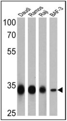
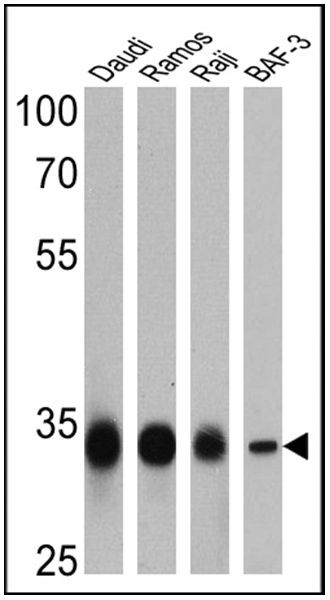
- Western blot analysis of CD20 was performed by loading 25 µg of Daudi (lane 1), Ramos (lane 2), Raji (lane 3) and BAF-3 (lane 4) cell lysates onto an SDS polyacrylamide gel. Proteins were transferred to a PVDF membrane and blocked at 4ºC overnight. The membrane was probed with a CD20 polyclonal antibody (Product # PA5-16701) at a dilution of 1:10,000 (Daudi and Ramos) and 1:200 (Raji and BAF-3) overnight at 4°C, washed in TBST, and probed with an HRP-conjugated secondary antibody for 1 hr at room temperature in the dark. Chemiluminescent detection was performed using Pierce ECL Plus Western Blotting Substrate (Product # 32132). Results show a band at ~33 kDa.
- Submitted by
- Invitrogen Antibodies (provider)
- Main image

- Experimental details
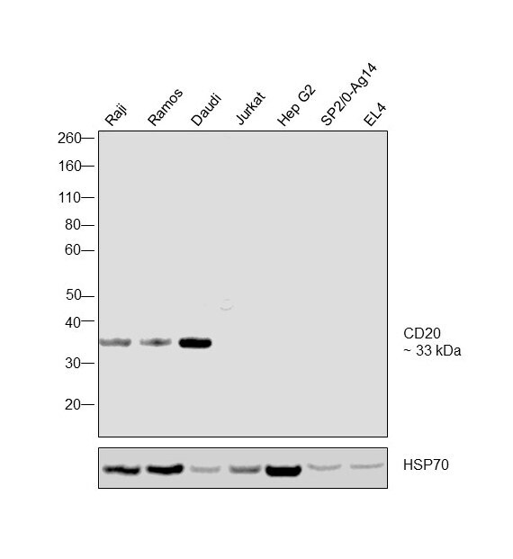
- Western blot was performed using Anti-CD20 Polyclonal Antibody, (Product # PA5-16701) and 33 kDa band corresponding to CD20 was observed in the cell lines tested except in Jurkat, Hep G2 which are low expressing models. Mouse cell lines showed no reactivity. Membrane enriched cell extracts (40 µg lysate) of Raji (Lane 1), Ramos (Lane 2), Daudi (Lane 3), Jurkat (Lane 4), Hep G2 (Lane 5), SP2/0-Ag14 (Lane 6) and EL4 (Lane 7) were electrophoresed using Novex® NuPAGE® 4-12 % Bis-Tris gel (Product # NP0321BOX). Resolved proteins were then transferred onto a nitrocellulose membrane (Product # IB23001) by iBlot® 2 Dry Blotting System (Product # IB21001). The blots were probed with the primary antibody (1:2000 dilution) and detected by chemiluminescence with Goat anti-Rabbit IgG (Heavy Chain), Superclonal™ Recombinant Secondary Antibody, HRP conjugate (Product # A27036, 1:4000 dilution) using the iBright FL 1000 (Product # A32752). Chemiluminescent detection was performed using Novex® ECL Chemiluminescent Substrate Reagent Kit (Product # WP20005).
Supportive validation
- Submitted by
- Invitrogen Antibodies (provider)
- Main image
- Experimental details
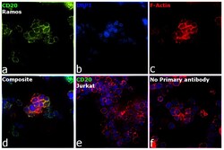
- Immunofluorescence analysis of CD20 was performed using 70% confluent log phase Ramos cells. The cells were fixed with 4% paraformaldehyde for 10 minutes, permeabilized with 0.1% Triton™ X-100 for 15 minutes, and blocked with 2% BSA for 1 hour at room temperature. The cells were labeled with CD20 Polyclonal Antibody (Product # PA5-16701) at 1:100 dilution in 0.1% BSA, incubated at 4 degree celsius overnight and then with Donkey anti-Rabbit IgG (H+L) Highly Cross-Adsorbed Secondary Antibody, Alexa Fluor Plus 488 (Product # A32790) at a dilution of 1:2000 for 45 minutes at room temperature (Panel a: green). Nuclei (Panel b: blue) were stained with SlowFade® Gold Antifade Mountant with DAPI (Product # S36938). F-actin (Panel c: red) was stained with Rhodamine Phalloidin (Product # R415, 1:300). Panel d represents the merged image showing localization in plasma membrane. Panel e represents negative cell line Jurkat with no expression. Panel f represents control cells with no primary antibody to assess background. The images were captured at 60X magnification.

- Submitted by
- Invitrogen Antibodies (provider)
- Main image

- Experimental details
- Immunofluorescence analysis of CD20 was performed using 70% confluent log phase Ramos cells. The cells were fixed with 4% paraformaldehyde for 10 minutes, permeabilized with 0.1% Triton™ X-100 for 15 minutes, and blocked with 2% BSA for 1 hour at room temperature. The cells were labeled with CD20 Polyclonal Antibody (Product # PA5-16701) at 1:100 dilution in 0.1% BSA, incubated at 4 degree celsius overnight and then with Donkey anti-Rabbit IgG (H+L) Highly Cross-Adsorbed Secondary Antibody, Alexa Fluor Plus 488 (Product # A32790) at a dilution of 1:2000 for 45 minutes at room temperature (Panel a: green). Nuclei (Panel b: blue) were stained with SlowFade® Gold Antifade Mountant with DAPI (Product # S36938). F-actin (Panel c: red) was stained with Rhodamine Phalloidin (Product # R415, 1:300). Panel d represents the merged image showing localization in plasma membrane. Panel e represents negative cell line Jurkat with no expression. Panel f represents control cells with no primary antibody to assess background. The images were captured at 60X magnification.

Supportive validation
- Submitted by
- Invitrogen Antibodies (provider)
- Main image
- Experimental details
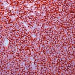
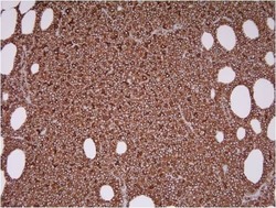
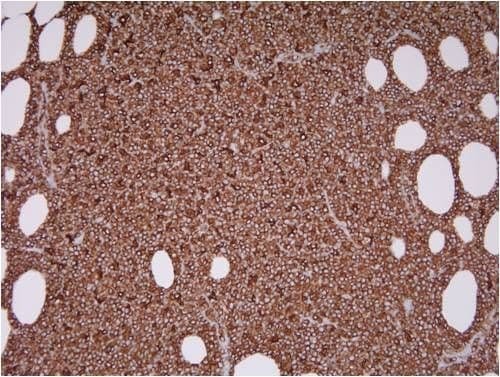
- Formalin-fixed, paraffin-embedded human tonsil stained with CD20 using peroxidase-conjugate and AEC. Note cell membrane staining of B lymphocytes

- Submitted by
- Invitrogen Antibodies (provider)
- Main image
- Experimental details
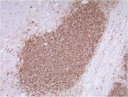
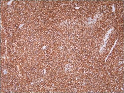
- Immunohistochemistry was performed on formalin-fixed, paraffin-embedded canine lymph node using an automated staining platform. Following deparaffinization, antigen retrieval was performed using CC1 retrieval solution for 30 minutes followed by 32 minute primary antibody incubation with rabbit anti-CD20 polyclonal antibody (Product # PA5-16701) at a dilution of 1:100. Detection was performed using an indirect biotin-streptavidin system and a DAB Detection kit. Data courtesy of the Innovators Program.
- Submitted by
- Invitrogen Antibodies (provider)
- Main image

- Experimental details
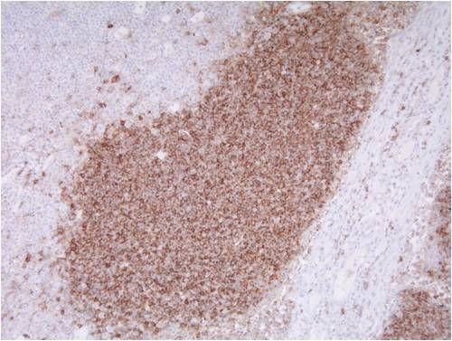
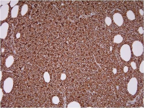
- Immunohistochemistry was performed on formalin-fixed, paraffin-embedded feline B cell lymphoma using an automated staining platform. Following deparaffinization, antigen retrieval was performed using CC1 retrieval solution for 30 minutes followed by 32 minute primary antibody incubation with rabbit anti-CD20 polyclonal antibody (Product # PA5-16701) at a dilution of 1:100. Detection was performed using an indirect biotin-streptavidin system and a DAB Detection kit. Data courtesy of the Innovators Program.
- Submitted by
- Invitrogen Antibodies (provider)
- Main image

- Experimental details
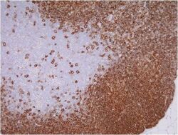
- Immunohistochemistry was performed on formalin-fixed, paraffin-embedded canine B cell lymphoma using an automated staining platform. Following deparaffinization, antigen retrieval was performed using CC1 retrieval solution for 30 minutes followed by 32 minute primary antibody incubation with rabbit anti-CD20 polyclonal antibody (Product # PA5-16701) at a dilution of 1:100. Detection was performed using an indirect biotin-streptavidin system and a DAB Detection kit. Data courtesy of the Innovators Program.
- Submitted by
- Invitrogen Antibodies (provider)
- Main image
- Experimental details
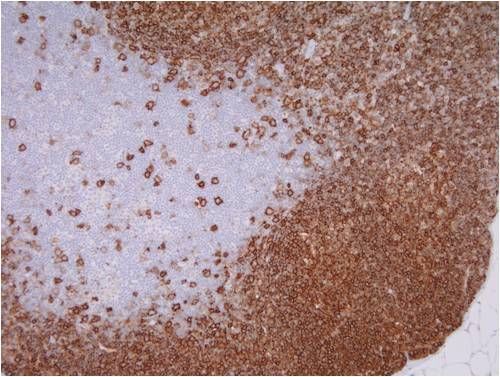
- Immunohistochemistry was performed on formalin-fixed, paraffin-embedded feline lymph node using an automated staining platform. Following deparaffinization, antigen retrieval was performed using CC1 retrieval solution for 30 minutes followed by 32 minute primary antibody incubation with rabbit anti-CD20 polyclonal antibody (Product # PA5-16701) at a dilution of 1:100. Detection was performed using an indirect biotin-streptavidin system and a DAB Detection kit. Data courtesy of the Innovators Program.
- Submitted by
- Invitrogen Antibodies (provider)
- Main image
- Experimental details
- Formalin-fixed, paraffin-embedded human tonsil stained with CD20 using peroxidase-conjugate and AEC. Note cell membrane staining of B lymphocytes
- Submitted by
- Invitrogen Antibodies (provider)
- Main image

- Experimental details
- Immunohistochemistry was performed on formalin-fixed, paraffin-embedded canine lymph node using an automated staining platform. Following deparaffinization, antigen retrieval was performed using CC1 retrieval solution for 30 minutes followed by 32 minute primary antibody incubation with rabbit anti-CD20 polyclonal antibody (Product # PA5-16701) at a dilution of 1:100. Detection was performed using an indirect biotin-streptavidin system and a DAB Detection kit. Data courtesy of the Innovators Program.

- Submitted by
- Invitrogen Antibodies (provider)
- Main image
- Experimental details
- Immunohistochemistry was performed on formalin-fixed, paraffin-embedded canine B cell lymphoma using an automated staining platform. Following deparaffinization, antigen retrieval was performed using CC1 retrieval solution for 30 minutes followed by 32 minute primary antibody incubation with rabbit anti-CD20 polyclonal antibody (Product # PA5-16701) at a dilution of 1:100. Detection was performed using an indirect biotin-streptavidin system and a DAB Detection kit. Data courtesy of the Innovators Program.
Supportive validation
- Submitted by
- Invitrogen Antibodies (provider)
- Main image

- Experimental details
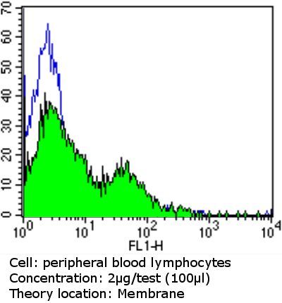
- Flow cytometry analysis of CD20 in peripheral blood mononuclear cells compared to an isotype control (blue). Human blood was collected, combined with a hydrophilic polysaccharide, centrifuged, transferred to a conical tube and washed with PBS. 50 ul of cell solution was added to each tube at a dilution of 2x10^7 cells/ml, followed by the addition of 50 ul of isotype control and primary antibody (Product # PA5-16701) at a dilution of 2 ug/test. Cells were incubated for 30 min at 4°C and washed with a cell buffer, followed by incubation with a DyLight 488-conjugated goat anti-rabbit IgG (H+L) secondary for 30 min at 4°C in the dark. FACS analysis was performed using 400 ul of cell buffer.
- Submitted by
- Invitrogen Antibodies (provider)
- Main image
- Experimental details
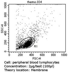
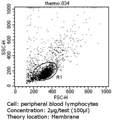
- Flow cytometry analysis of CD20 in peripheral blood mononuclear cells compared to an isotype control (blue). Human blood was collected, combined with a hydrophilic polysaccharide, centrifuged, transferred to a conical tube and washed with PBS. 50 ul of cell solution was added to each tube at a dilution of 2x10^7 cells/ml, followed by the addition of 50 ul of isotype control and primary antibody (Product # PA5-16701) at a dilution of 2 ug/test. Cells were incubated for 30 min at 4°C and washed with a cell buffer, followed by incubation with a DyLight 488-conjugated goat anti-rabbit IgG (H+L) secondary for 30 min at 4°C in the dark. FACS analysis was performed using 400 ul of cell buffer.
Supportive validation
- Submitted by
- Invitrogen Antibodies (provider)
- Main image
- Experimental details
- NULL
- Submitted by
- Invitrogen Antibodies (provider)
- Main image
- Experimental details
- NULL
- Submitted by
- Invitrogen Antibodies (provider)
- Main image
- Experimental details
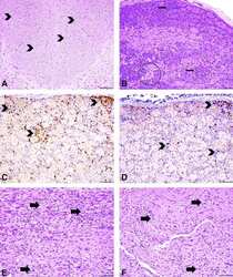
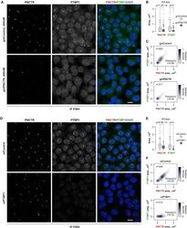
- Figure 3 Representative CNS and PNS Histopathologic Findings in Rhesus Macaques Injected with Suboccipital AAV9.hIDUA (A-C) The majority of animals had minimal to mild neuronal cell body degeneration in the DRG characterized by neurodegeneration (arrow, A), satellitosis, and mononuclear cell infiltrates that surrounded and invaded neuronal cell bodies (arrows, B). A normal DRG from a vehicle control animal is represented for comparison (C). (D-F) All animals from test article-treated groups had an axonopathy in the dorsal white matter tracts of the spinal cord that was bilateral and characterized by dilated myelin sheaths with and without myelomacrophages (arrows), consistent with axonal degeneration. An unremarkable section from the vehicle control group is shown for comparison (F). H&E staining was used. (G) Individual cumulative scores defined as the sum of cervical, thoracic, and lumbar segments scores with 1 as minimal, 2 as mild, 3 as moderate, 4 as marked, and 5 as severe; no animal had a score higher than 3 in any segment analyzed. Error bars represent SDs. (H) Immunofluorescence staining of the DRG using anti-hIDUA antibodies (red), anti-CD20 or anti-CD3 antibodies (green), and DAPI (nuclear counterstaining). Mononuclear cell infiltrates were predominantly composed of CD3 + T cells (B) and CD20 + B cells (C). Scale bars, 200 mum (A-C) and 100 mum (D-F).
- Submitted by
- Invitrogen Antibodies (provider)
- Main image
- Experimental details
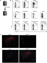
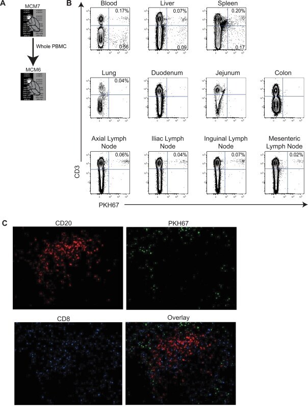
- Figure 6 Lymphocytes traffic to peripheral lymphoid tissues after adoptive transfer. A Diagram displaying the transfer protocol. B Donor bulk PBMC were transferred from MCM7 into MCM6. MCM6 was euthanized 24 hours post transfer and its tissues were processed. Cells selectively trafficked to different peripheral lymphoid organs post-transfer. C Immunohistochemical staining of a lymph node section showing PKH67+ cells (green), CD20 antibody staining (red) and CD8 antibody staining (blue) collected with a confocal microscope at 200X.
- Submitted by
- Invitrogen Antibodies (provider)
- Main image
- Experimental details
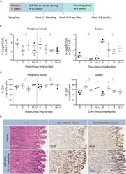
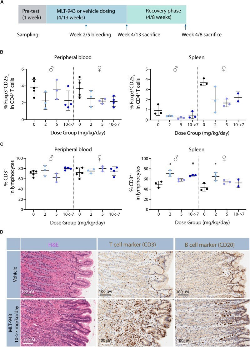
- FIGURE 6 Prolonged pharmacological MALT1 protease inhibition leads to reduced Tregs in dogs. (A) Outline of 4-week and 13-week GLP toxicity studies including pre-test, treatment and recovery phases. (B) Frequency of Foxp3 + CD25 + Tregs and (C) CD3 + T cells in blood and spleen in male and female beagle dogs determined by FACS at time of necropsy (day 86 or recovery day 55). Each dot represents an individual animal. Lines depict mean +- SD. Statistical difference was determined using one-way ANOVA with follow up for significance by multiple comparison tests with Sidak's correction,* p < 0.05. (D) Histological alterations in stomach of beagle dogs treated with MLT-943 for up to 13 weeks. All animals were females and belonged to either the vehicle or the 10-7 mg/kg/day group. Sections taken at necropsy day 86 and stained with H&E (left panel), anti-CD3 (middle panel), or anti-CD20 (right panel).
- Submitted by
- Invitrogen Antibodies (provider)
- Main image
- Experimental details
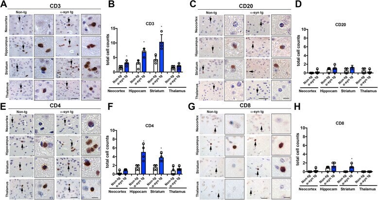
- Fig. 1 Immunohistochemical analysis of lymphoid cells in the brains of -synuclein transgenic mice. a Representative bright field light microscopy images from the neocortex, hippocampus, striatum, and thalamus of non-tg and -syn tg mice immunostained with an antibody against CD3 (general T cell marker). b Computer-based image analysis showing significant increase of CD3 positive cell numbers in neocortex, hippocampus, and striatum of -syn tg mice. c Representative bright field light microscopy images from the neocortex, hippocampus, striatum, and thalamus of non-tg and -syn tg mice immunostained with an antibody against CD20 (general B cell marker). d Computer-based image analysis showing comparable numbers of CD20 positive cell in the brains of non-tg and -syn tg mice. e Representative bright field light microscopy images from the neocortex, hippocampus, striatum, and thalamus of non-tg and -syn tg mice immunostained with an antibody against CD4 (helper T cell marker). f Computer-based image analysis showing significant increase of CD4 positive cell numbers in neocortex, hippocampus and striatum of -syn tg mice. g Representative bright field light microscopy images from the neocortex, hippocampus, striatum, and thalamus of non-tg and -syn tg mice immunostained with an antibody against CD8 (cytotoxic T cell marker). h Computer-based image analysis showing significant increase of CD8 positive cell numbers in neocortex, hippocampus, and striatum of -syn tg mice. Scale bars = 40
- Submitted by
- Invitrogen Antibodies (provider)
- Main image
- Experimental details
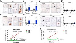
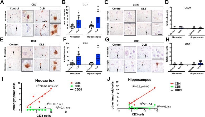
- Fig. 4 Immunohistochemical analysis of lymphoid cells in the brains of DLB cases. a Representative bright field light microscopy images from the neocortex and hippocampus of control and DLB cases immunostained with an antibody against CD3 (general T cell marker). b Computer-based image analysis showing significant increase of CD3 positive cell numbers in neocortex and hippocampus in DLB. c Representative bright field light microscopy images from the neocortex and hippocampus of control and DLB immunostained with an antibody against CD20 (general B cell marker). d Computer-based image analysis showing very few or none CD20-positive cell in the brains. e Representative bright field light microscopy images from the neocortex and hippocampus of control and DLB cases, immunostained with an antibody against CD4 (helper T cell marker). f Computer-based image analysis showing significant increase of CD4-positive cell numbers in neocortex and hippocampus in DLB. g Representative bright field light microscopy images from the neocortex and hippocampus in control and DLB immunostained with an antibody against CD8 (cytotoxic T cell marker). h Computer-based image analysis showing few CD8-positive cells in human brains. i , j Linear regression analysis between CD3 and CD4, CD8, and CD20 in the neocortex and hippocampus. Scale bars = 40 mum (low magnification) and 10 mum (high magnification). Control and DLB ( n = 8 per group). Statistical significance determined by unpaired t test; * p
- Submitted by
- Invitrogen Antibodies (provider)
- Main image
- Experimental details
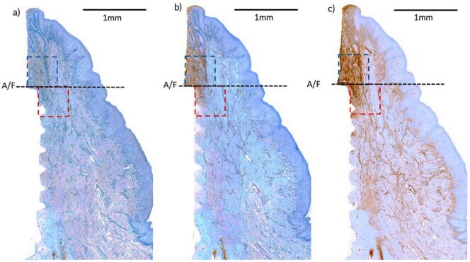
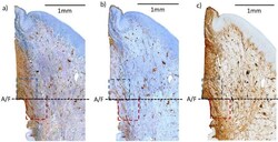
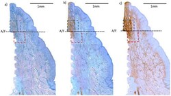
- Figure 4 Immunohistochemical (IHC) stained histological slides of different types of primary antibody--( a ) CD3, ( b ) CD20, ( c ) IgG antibody. A/F--abutment-fixture junction; upper (blue) and lower (maroon) region of interest (ROI) selected for cell marker intensity analysis.
- Submitted by
- Invitrogen Antibodies (provider)
- Main image
- Experimental details
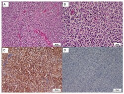
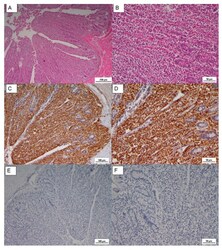
- Figure 2 ( A ) Microscopic lesion of tumor mass, uniform proliferation of lymphoblast and small to medium lymphocyte, HE, Scale bar = 50 um; ( B ) HE, Scale bar = 20 um; ( C ) IHC CD3, diffuse positive staining, Scale bar = 50 um; ( D ) IHC CD20, negative staining, Scale bar = 50 um.

- Submitted by
- Invitrogen Antibodies (provider)
- Main image
- Experimental details
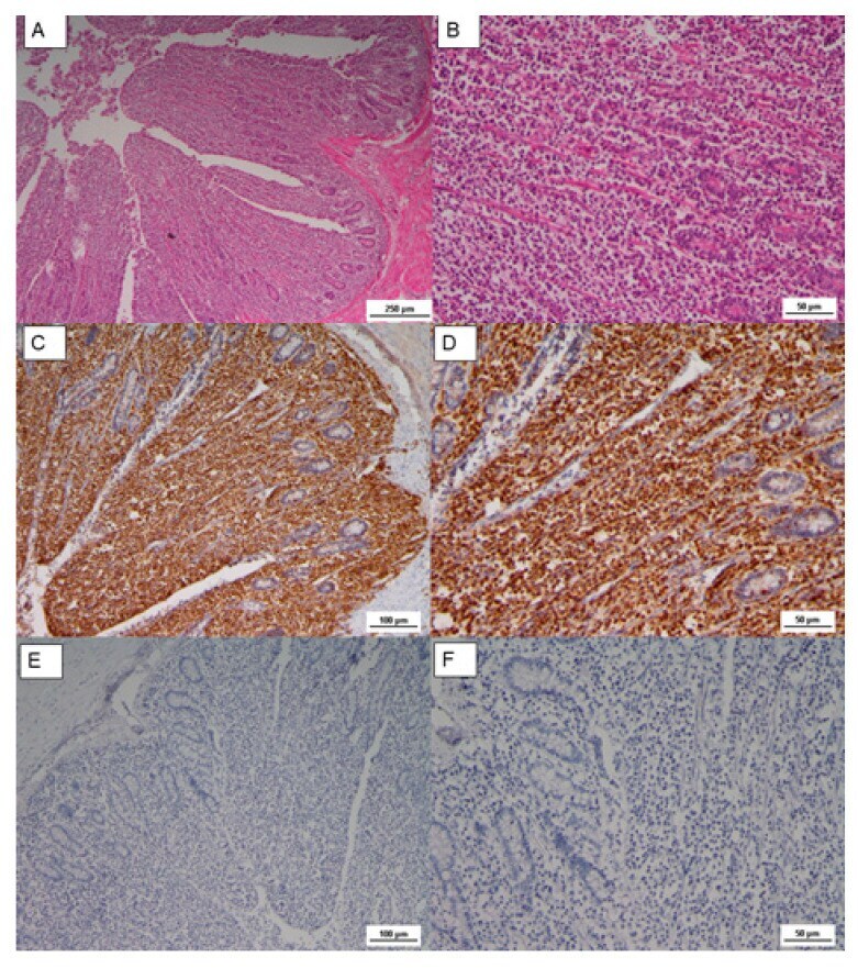
- Figure 3 ( A ) Microscopic lesion of cecal tonsils, loss of intestine structure, caused by neoplastic proliferation and massive infiltration of lamina propria, HE, Scale bar = 250 um; ( B ) Lymphoblasts with pleomorphic nuclei, and small to medium in size lymphocytes, HE Scale bar = 50 um; ( C ) IHC CD3, diffuse positive staining, Scale bar = 100 um; ( D ) IHC CD3 Scale bar= 50 um; ( E ) IHC CD20, negative staining, Scale bar = 1000 um; ( F ) IHC CD20, Scale bar = 50 um.
- Submitted by
- Invitrogen Antibodies (provider)
- Main image
- Experimental details
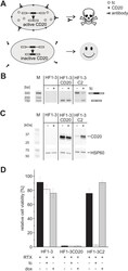
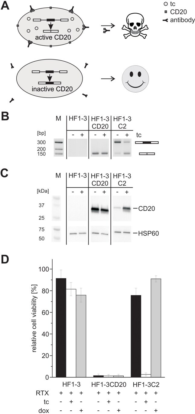
- Figure 6. The tc aptamer controlled CD20 expression. ( A ) Schematic representation of the suicide system. When treated with the therapeutic antibody rituximab (RTX), cell death was induced in cells expressing CD20. As a positive control, CD20 was stably integrated into HeLa HF1-3 cells (cell line HF1-3CD20). The aptamer-controlled exon 2 along with the neighboring introns from construct L2 was inserted into the CD20 coding region, which resulted in the cell line HF1-3C2. In the presence of tc, exon 2 was skipped, the coding sequence of CD20 was restored, and apoptosis was triggered. Without tc, exon 2 was retained, which destroyed the CD20 reading frame and resulted in cell survival. ( B, C ) Cell lines were incubated with and without 50 muM tc for 2 days. Afterwards, RT-PCR and a western blot were performed. Primers used for RT-PCR bind to the coding sequence of CD20, which spans the alternatively spliced exon 2. HSP60 was used as a loading control for western blot. ( D ) Viability assay. The cell lines were incubated with and without RTX and 50 muM tc or doxycycline (dox) for 3 days. Afterwards, cell viability was analyzed by an AlamarBlue (r) assay. Error bars represent the standard deviation from mean values. For each cell line, the value of untreated cells was set to 100. The values of untreated cells are shown in Supplementary Figure S6 .
- Submitted by
- Invitrogen Antibodies (provider)
- Main image
- Experimental details
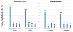
- Figure 1 Intra-species differences in the numbers of macrophages (based on Iba1 immunostaining), plasma cells (lambda chain), T lymphocytes (CD3) and B lymphocytes (CD20) in skin mange lesions from wild carnivores and ruminants. For each animal species, the same letters above different rectangular bars indicates significant differences between means. For wolf: (a, b, c) p < 0.001. For fox: (a, b, c, d, e) p < 0.05. For chamois: (a, b) p < 0.05. For red deer: (a, b, c) p < 0.001. A Tukey test for multiple comparisons was applied for statistical analysis.

- Submitted by
- Invitrogen Antibodies (provider)
- Main image
- Experimental details
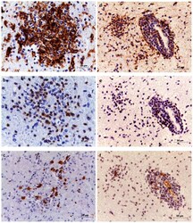
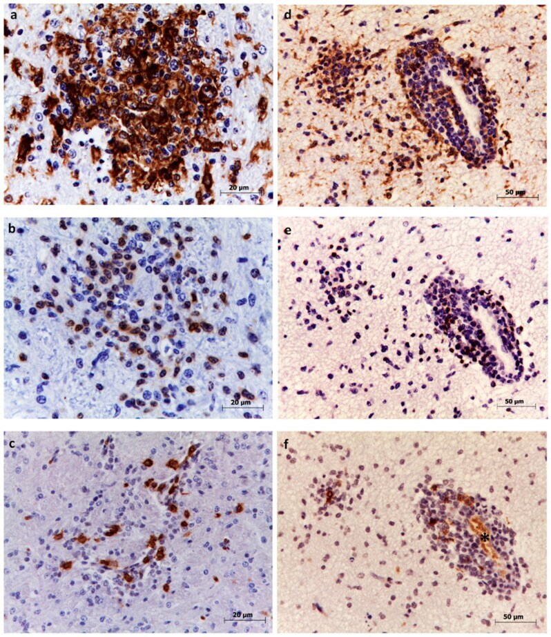
- Figure 2 Immunohistochemistry to detect microglia, T lymphocytes, and B lymphocytes in thalamus ( a - c ) and hippocampus ( d - f ). Microglia were identified based on labeling with anti-Iba1 antibodies. Abundant cells are visible within glial foci ( a ) and in the perivascular infiltrate ( d ). T lymphocytes were identified based on labeling with anti-CD3 antibodies. Moderate numbers are visible in the glial focus ( b ) and in the perivascular cuff ( e ). B lymphocytes were identified based on labeling with anti-CD20 antibodies. Few numbers are visible within the glial focus ( c ) and perivascular infiltrate ( f ). Erythrocytes were also immunolabelled (asterisk) but were not included in the counting.
- Submitted by
- Invitrogen Antibodies (provider)
- Main image

- Experimental details
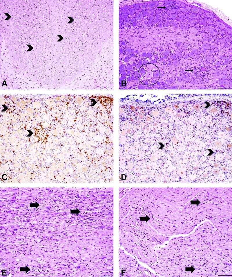
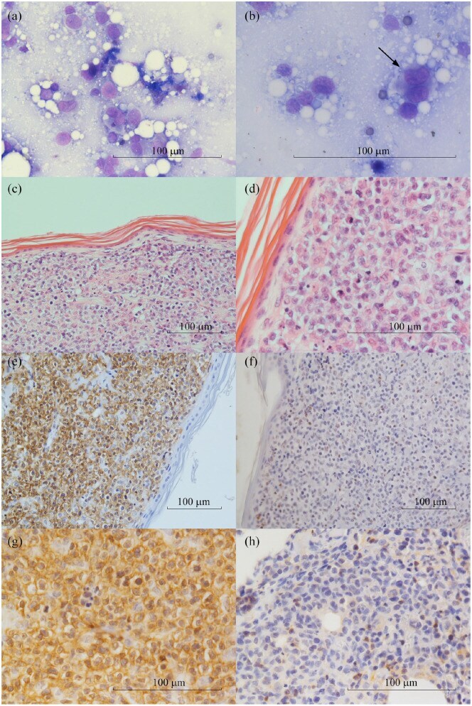
- Figure 2 Feline cutaneous non-epitheliotropic B-cell lymphoma. (a) A population of medium and large round elements has been highlighted, with abundant cytoplasm and a round-to-cleaved nucleus with prominent and irregular nucleolus (May-Grunwald-Giemsa [MGG], x 40). (b) Several binucleated (arrow) cells can be seen (MGG, x 60). (c) Medium and large round neoplastic cells infiltrate extensively the superficial and deep dermis (haematoxylin and eosin [H&E], x 20). (d) There was no evidence of epitheliotropism in the neoplastic cells (H&E, x 40). (e-g) Neoplastic cells show strong positivity to CD20 marker (immunohistochemistry [IHC], x 20, x 40). (f-h) Few cells show positivity to marker CD3 (IHC, x 20, x 40)
- Submitted by
- Invitrogen Antibodies (provider)
- Main image
- Experimental details
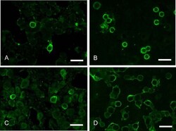
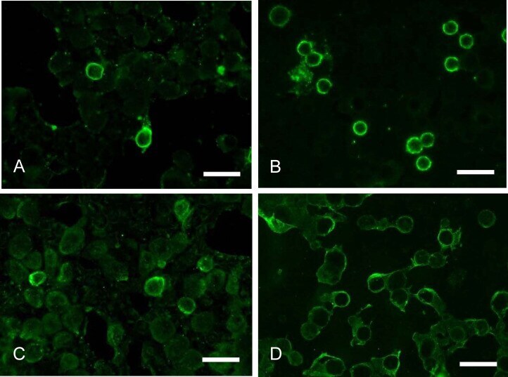
- Fig. 2. Comparison of the rapid multiple immunofluorescence (RMIF) CD20/CD3 and RMIF CD79alpha/CD3 methods. A & B: Detection of CD20. C & D: Detection of CD79alpha. A & C: Fine needle biopsy samples from the lymph node of a dog with T-cell lymphoma. B & D: Sediment smear from the chylous effusion of a cat. In the RMIF CD20/CD3 samples, green signals for CD20 were observed in the margins of certain lymphocytes, and negative reactions were clearly observable in most lymphocytes. In the RIMF CD79alpha/CD3 samples, the weak, nonspecific green signals were detected in multiple lymphocytes. In all panels, a fluorescence filter adapted to Alexa 488 was used. Scale bars=10 um.
- Submitted by
- Invitrogen Antibodies (provider)
- Main image
- Experimental details
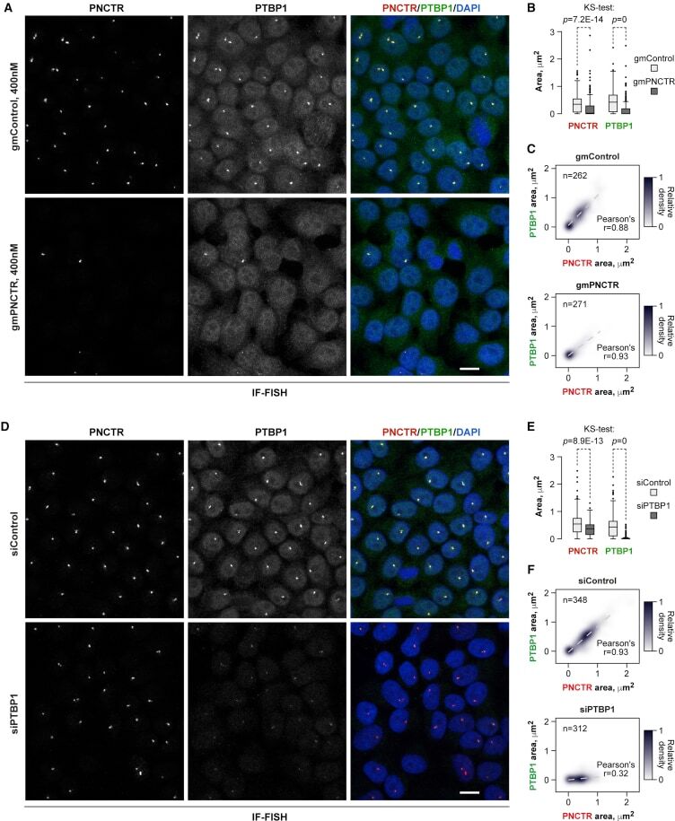
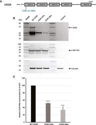
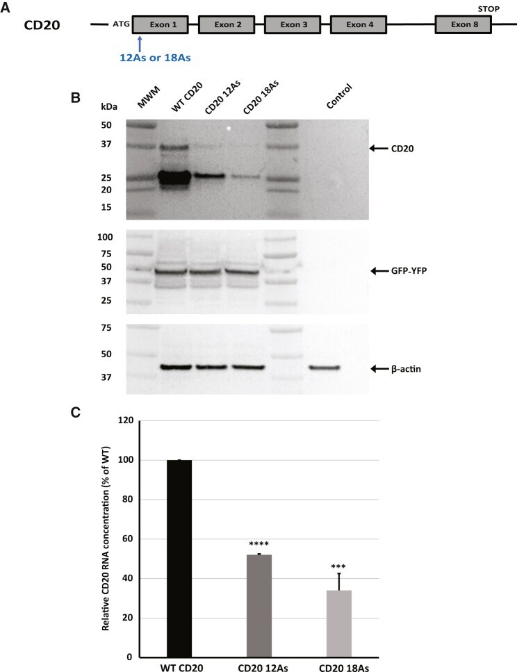
- Figure 1 Design and effect of poly(A) track insertion on CD20 stability and expression (A) Scheme representative of poly(A) insertions and location of insertions for CD20 reporter constructs used in this study. Poly(A) insertions in the CD20 coding sequence were placed proximal to the ATG start codon. A schematic of the CD20 gene with the start codon (ATG), termination codon (STOP), and exons and insertion sites is also shown. (B) Western blot analysis of CD20 constructs expressed transiently in CHO cells. An equal amount of total protein was used for analysis. GFP-YFP and beta-actin were used as transfection and loading controls, respectively, with expression of both proteins detected using specific antibodies. CD20 was detected with a CD20-specfic antibody. Arrows indicate the position of full-length CD20, GFP-YFP, and beta-actin proteins. (C) mRNA levels of CD20 constructs were measured by qRT-PCR. CD20 expression was normalized to beta-actin. Relative mRNA levels for both constructs (12As and 18As) are presented as percentages of WT CD20. Error bars represent mean +- SD (n = 3). p values represent significance (*p
- Submitted by
- Invitrogen Antibodies (provider)
- Main image
- Experimental details
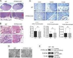
- FIGURE 2 Sections of immunostaining with a polyclonal anti-CD20 + mab of canine mammary sections from animals affected by breast cancer. (A) CD20 + Lymphocyte infiltrating tumor sample (20X) in a G1 sample; (B) CD20 + Lymphocyte infiltrating tumor sample (20X) in a G2 sample; (C) CD20 + Lymphocyte infiltrating tumor sample (20X) in a G3 sample.

- Submitted by
- Invitrogen Antibodies (provider)
- Main image
- Experimental details
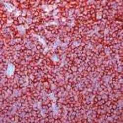
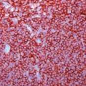
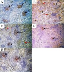
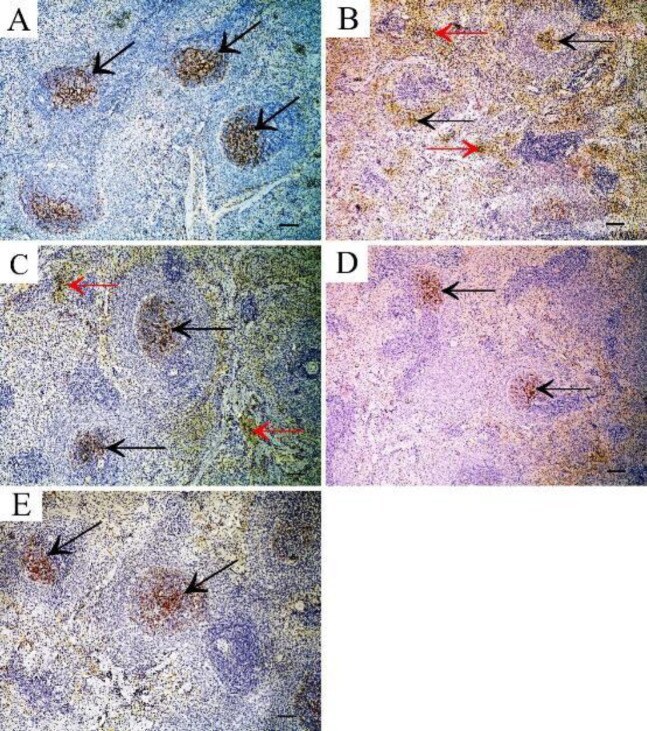
- Figure 8 CD20 immunostained sections in the spleen showing A: control group shows CD20+ B cells appearing mainly in lymphatic follicles of white pulp (arrow). B: TAA-treated group showing increased CD20+ B cells in lymphatic follicles of white pulp (black arrow) and in red pulp (red arrow). C: rapamycin-treated group showing CD20+ B cells in lymphatic follicles of white pulp (black arrow) and few in the red pulp (red arrow). D: Filgrastim-treated group showing CD20+ B cells in lymphatic follicles of white pulp (arrow). E: combined filgrastim and rapamycin-treated group showing CD20+ B cells in lymphatic follicles of white pulp (arrow) (Immunoperoxidase technique for CD20 x 10, scale bar 50 um) TTA: thioacetamide
- Submitted by
- Invitrogen Antibodies (provider)
- Main image
- Experimental details
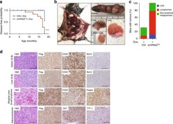
- Fig. 6 UVRAG FS promotes spontaneous tumorigenesis in mice. a Kaplan-Meier plot of time to development of any malignancy in Dox-treated iUVRAG FS mice ( n = 25) versus Dox-treated control mice ( n = 25). Malignancy is determined by complete histologic survey of all major internal organs. *** P < 0.001 (Log-rank test). b Representative images of DLBCL in different organs that developed in Dox-treated iUVRAG FS mice. c Percentage of control ( n = 16) and UVRAG FS -expressing ( n = 12) mice with spontaneous tumors, including lymphoproliferative disease (LPD), lymphomas, and non-lymphoid malignancies. d H&E and IHC analysis of the most frequently observed neoplastic lesions from Dox-treated iUVRAG FS mice using the antibodies as indicated. Scale bars, 100 mum.

- Submitted by
- Invitrogen Antibodies (provider)
- Main image
- Experimental details
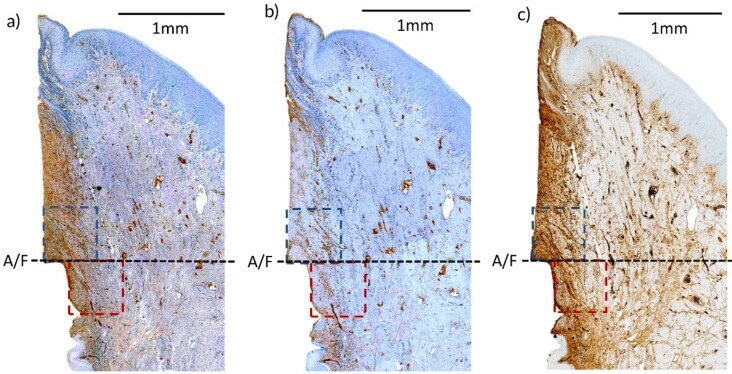
- Figure 3 IHC staining of an implant using fracture technique (20x magnification). ( a ) CD3; ( b ) CD20; ( c ) IgG. Upper (Blue) and lower (Maroon) region of interest are labelled. A/F--abutment-fixture junction.