Explore
 Validate
 Learn
Learn Immunohistochemistry
Antibody data
- Antibody Data
- Antigen structure
- References [1]
- Comments [0]
- Validations
- Immunohistochemistry [1]
- Flow cytometry [1]
Submit
Validation data
Reference
Comment
Report error
- Product number
- MAB4225 - Provider product page

- Provider
- R&D Systems
- Product name
- Human CD20 Antibody
- Antibody type
- Monoclonal
- Description
- Protein A or G purified from hybridoma culture supernatant. Detects human CD20. Stains human CD20 transfectants but not irrelevant transfectants.
- Reactivity
- Human
- Host
- Mouse
- Conjugate
- Unconjugated
- Antigen sequence
P11836- Isotype
- IgG
- Antibody clone number
- 396444
- Vial size
- 100 ug
- Concentration
- LYOPH
- Storage
- Use a manual defrost freezer and avoid repeated freeze-thaw cycles. 12 months from date of receipt, -20 to -70 °C as supplied. 1 month, 2 to 8 °C under sterile conditions after reconstitution. 6 months, -20 to -70 °C under sterile conditions after reconstitution.
Submitted references Preclinical Development of a Bispecific Antibody that Safely and Effectively Targets CD19 and CD47 for the Treatment of B-Cell Lymphoma and Leukemia.
Buatois V, Johnson Z, Salgado-Pires S, Papaioannou A, Hatterer E, Chauchet X, Richard F, Barba L, Daubeuf B, Cons L, Broyer L, D'Asaro M, Matthes T, LeGallou S, Fest T, Tarte K, Clarke Hinojosa RK, Genescà Ferrer E, Ribera JM, Dey A, Bailey K, Fielding AK, Eissenberg L, Ritchey J, Rettig M, DiPersio JF, Kosco-Vilbois MH, Masternak K, Fischer N, Shang L, Ferlin WG
Molecular cancer therapeutics 2018 Aug;17(8):1739-1751
Molecular cancer therapeutics 2018 Aug;17(8):1739-1751
No comments: Submit comment
Supportive validation
- Submitted by
- R&D Systems (provider)
- Main image
- Experimental details
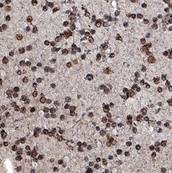
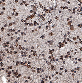
- CD20 in Human Leukemia. CD20 was detected in immersion fixed paraffin-embedded sections of human leukemia using Mouse Anti-Human CD20 Monoclonal Antibody (Catalog # MAB4225) at 15 µg/mL for 1 hour at room temperature followed by incubation with the Anti-Mouse IgG VisUCyte™ HRP Polymer Antibody (Catalog # VC001). Tissue was stained using DAB (brown) and counterstained with hematoxylin (blue). Specific staining was localized to lymphocytes. View our protocol for IHC Staining with VisUCyte HRP Polymer Detection Reagents.
Supportive validation
- Submitted by
- R&D Systems (provider)
- Main image
- Experimental details
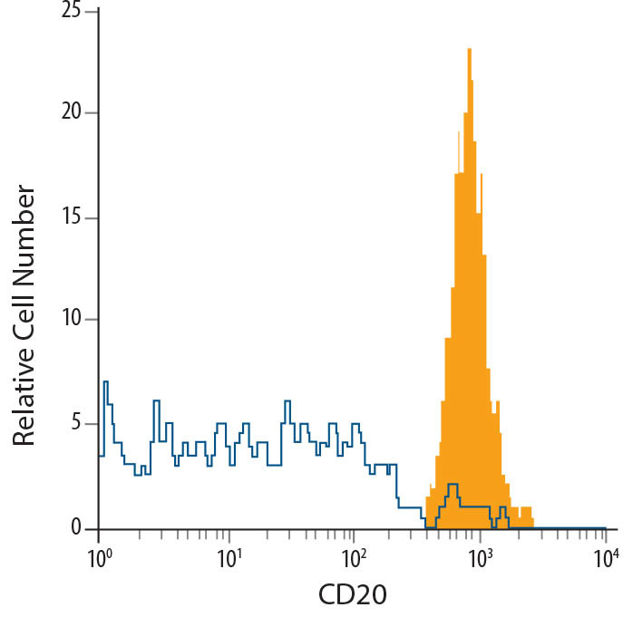
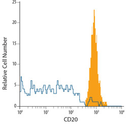
- Detection of CD20 in Human B Cells by Flow Cytometry. Human whole blood CD19+ B cells were stained with Mouse Anti-Human CD20 Monoclonal Antibody (Catalog # MAB4225, filled histogram) or isotype control antibody (Catalog # MAB002, open histo-gram), followed by Allophycocyanin-conjugated Anti-Mouse IgG F(ab')2 Secondary Antibody (Catalog # F0101B).