 Explore
 Validate
 Learn
Learn Western blot
 Immunocytochemistry
Antibody data
- Antibody Data
- Antigen structure
- References [3]
- Comments [0]
- Validations
- Immunocytochemistry [1]
- Immunohistochemistry [1]
Submit
Validation data
Reference
Comment
Report error
- Product number
- HPA014341 - Provider product page

- Provider
- Atlas Antibodies
- Proper citation
- Atlas Antibodies Cat#HPA014341, RRID:AB_1846265
- Product name
- Anti-MS4A1
- Antibody type
- Polyclonal
- Description
- Polyclonal Antibody against Human MS4A1, Gene description: membrane-spanning 4-domains, subfamily A, member 1, Alternative Gene Names: B1, Bp35, CD20, MS4A2, Validated applications: WB, IHC, ICC, Uniprot ID: P11836, Storage: Store at +4°C for short term storage. Long time storage is recommended at -20°C.
- Reactivity
- Human
- Host
- Rabbit
- Conjugate
- Unconjugated
- Isotype
- IgG
- Vial size
- 100 µl
- Concentration
- 0.1 mg/ml
- Storage
- Store at +4°C for short term storage. Long time storage is recommended at -20°C.
- Handling
- The antibody solution should be gently mixed before use.
Submitted references A lncRNA signature associated with tumor immune heterogeneity predicts distant metastasis in locoregionally advanced nasopharyngeal carcinoma
Single-cell transcriptomics reveals regulators underlying immune cell diversity and immune subtypes associated with prognosis in nasopharyngeal carcinoma
Arterial immune protein expression demonstrates the complexity of immune responses in Kawasaki disease arteritis
Liang Y, Zhang Y, Tan X, Qiao H, Liu S, Tang L, Mao Y, Chen L, Li W, Zhou G, Zhao Y, Li J, Li Q, Huang S, Gong S, Zheng Z, Li Z, Sun Y, Jiang W, Ma J, Li Y, Liu N
Nature Communications 2022;13(1)
Nature Communications 2022;13(1)
Single-cell transcriptomics reveals regulators underlying immune cell diversity and immune subtypes associated with prognosis in nasopharyngeal carcinoma
Chen Y, Yin J, Li W, Li H, Chen D, Zhang C, Lv J, Wang Y, Li X, Li J, Zhang P, Li Y, He Q, Yang X, Lei Y, Tang L, Zhou G, Mao Y, Wei C, Xiong K, Zhang H, Zhu S, Hou Y, Sun Y, Dean M, Amit I, Wu K, Kuang D, Li G, Liu N, Ma J
Cell Research 2020;30(11):1024-1042
Cell Research 2020;30(11):1024-1042
Arterial immune protein expression demonstrates the complexity of immune responses in Kawasaki disease arteritis
Cameron S, White S, Arrollo D, Shulman S, Rowley A
Clinical and Experimental Immunology 2017;190(2):244-250
Clinical and Experimental Immunology 2017;190(2):244-250
No comments: Submit comment
Supportive validation
- Submitted by
- Atlas Antibodies (provider)
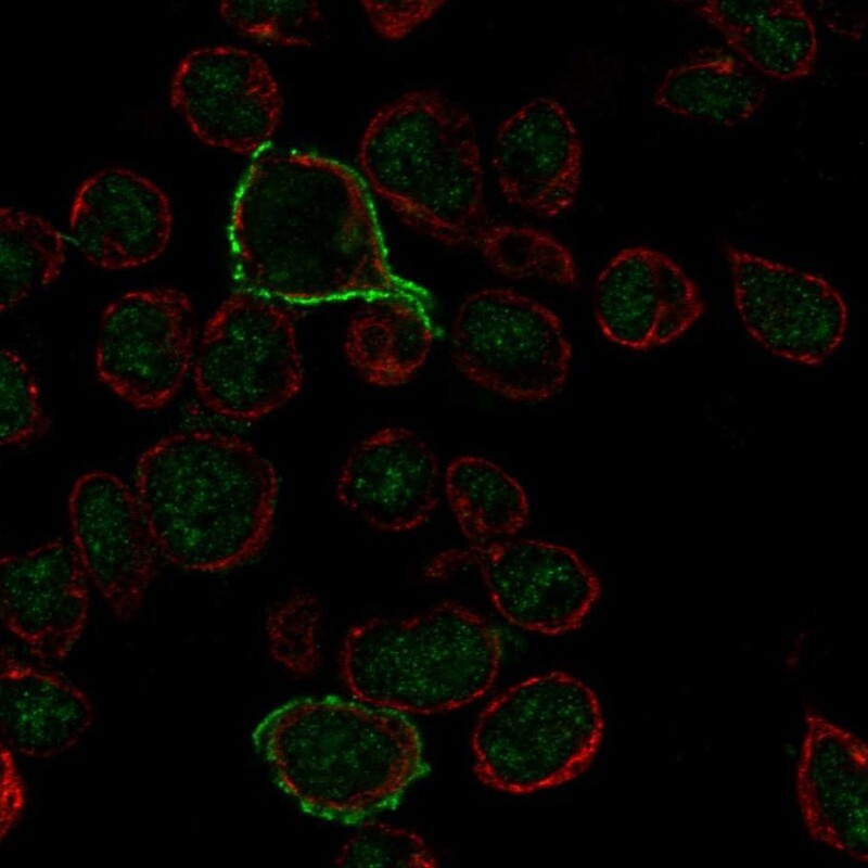
- Main image
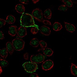
- Experimental details
- Immunofluorescent staining of human cell line REH shows localization to nucleus & plasma membrane.
- Sample type
- Human
Supportive validation
- Submitted by
- Atlas Antibodies (provider)
- Enhanced method
- Orthogonal validation
- Main image
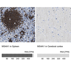
- Experimental details
- Immunohistochemistry analysis in human spleen and cerebral cortex tissues using HPA014341 antibody. Corresponding MS4A1 RNA-seq data are presented for the same tissues.
- Sample type
- Human
- Protocol
- Protocol
