Explore
 Validate
 Learn
Learn Western blot
 Immunocytochemistry
Antibody data
- Antibody Data
- Antigen structure
- References [4]
- Comments [0]
- Validations
- Immunocytochemistry [2]
- Immunohistochemistry [2]
- Flow cytometry [2]
- Other assay [2]
Submit
Validation data
Reference
Comment
Report error
- Product number
- PA5-85917 - Provider product page

- Provider
- Invitrogen Antibodies
- Product name
- CD34 Polyclonal Antibody
- Antibody type
- Polyclonal
- Antigen
- Synthetic peptide
- Reactivity
- Human, Mouse, Rat
- Host
- Rabbit
- Isotype
- IgG
- Vial size
- 100 μL
- Concentration
- 1 mg/mL
- Storage
- Store at 4°C short term. For long term storage, store at -20°C, avoiding freeze/thaw cycles.
Submitted references Propofol suppresses adipose-derived stem cell progression via PI3K/AKT-Wnt signaling pathway.
Over-expression of MEG3 promotes differentiation of bone marrow mesenchymal stem cells into chondrocytes by regulating miR-129-5p/RUNX1 axis.
Myotendinous junction adaptations to ladder-based resistance training: identification of a new telocyte niche.
Bone marrow mesenchymal stem cell-derived exosomes protect against myocardial infarction by promoting autophagy.
Yin G, Wang J, Zhong Y, Wu W
BMC anesthesiology 2022 Mar 9;22(1):65
BMC anesthesiology 2022 Mar 9;22(1):65
Over-expression of MEG3 promotes differentiation of bone marrow mesenchymal stem cells into chondrocytes by regulating miR-129-5p/RUNX1 axis.
Zhu J, Fu Q, Shao J, Peng J, Qian Q, Zhou Y, Chen Y
Cell cycle (Georgetown, Tex.) 2021 Jan;20(1):96-111
Cell cycle (Georgetown, Tex.) 2021 Jan;20(1):96-111
Myotendinous junction adaptations to ladder-based resistance training: identification of a new telocyte niche.
Pimentel Neto J, Rocha LC, Barbosa GK, Jacob CDS, Krause Neto W, Watanabe IS, Ciena AP
Scientific reports 2020 Aug 24;10(1):14124
Scientific reports 2020 Aug 24;10(1):14124
Bone marrow mesenchymal stem cell-derived exosomes protect against myocardial infarction by promoting autophagy.
Zou L, Ma X, Lin S, Wu B, Chen Y, Peng C
Experimental and therapeutic medicine 2019 Oct;18(4):2574-2582
Experimental and therapeutic medicine 2019 Oct;18(4):2574-2582
No comments: Submit comment
Supportive validation
- Submitted by
- Invitrogen Antibodies (provider)
- Main image
- Experimental details
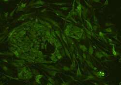
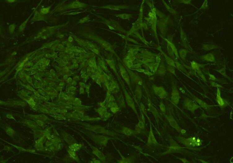
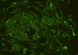
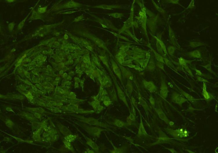
- Immunocytochemical analysis of CD34 in hES cells using a CD34 Polyclonal antibody (Product # PA5-85917) as seen in green. Cells were fixed in paraformaldehyde, permeabilised with 0.25% Triton X100/PBS.
- Submitted by
- Invitrogen Antibodies (provider)
- Main image
- Experimental details
- Immunocytochemical analysis of CD34 in hES cells using a CD34 Polyclonal antibody (Product # PA5-85917) as seen in green. Cells were fixed in paraformaldehyde, permeabilised with 0.25% Triton X100/PBS.
Supportive validation
- Submitted by
- Invitrogen Antibodies (provider)
- Main image

- Experimental details
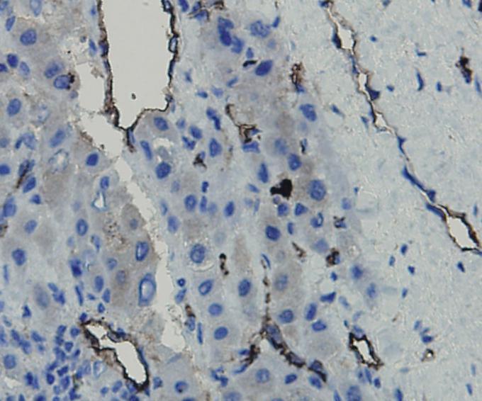
- Immunohistochemical analysis of CD34 of paraffin-embedded Human liver carcinoma tissue using a CD34 Polyclonal antibody (Product #PA5-85917). Counter stained with hematoxylin.
- Submitted by
- Invitrogen Antibodies (provider)
- Main image

- Experimental details
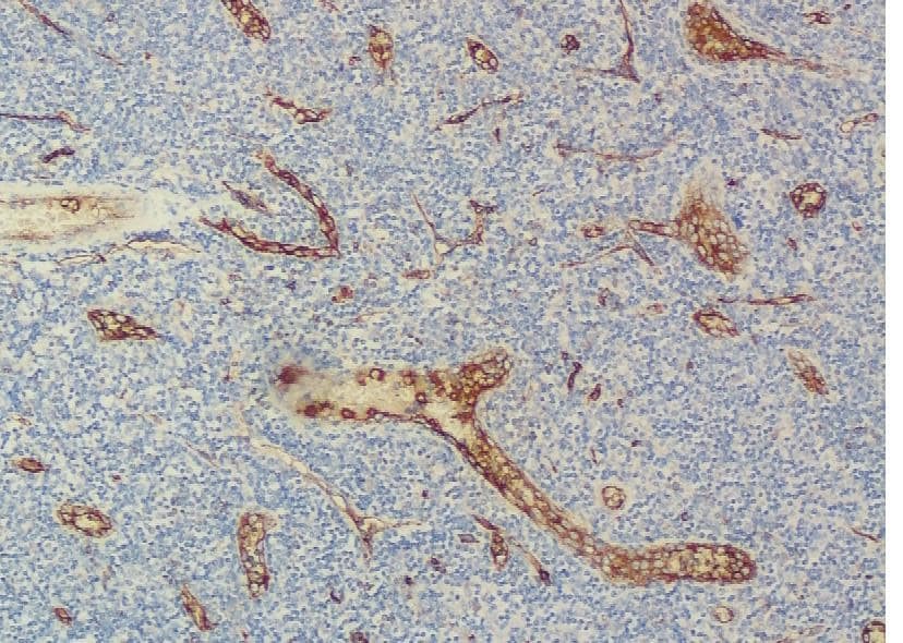
- Immunohistochemical analysis of CD34 of paraffin-embedded Human tonsil tissue using a CD34 Polyclonal antibody (Product #PA5-85917). Counter stained with hematoxylin.
Supportive validation
- Submitted by
- Invitrogen Antibodies (provider)
- Main image
- Experimental details
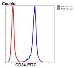
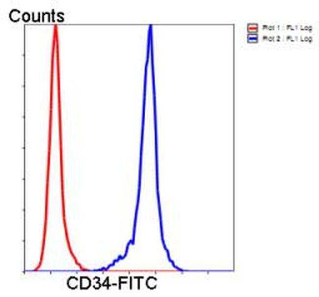
- Flow Cytometric analysis of CD34 in Jurkat cells using a CD34 Polyclonal Antibody (Product # PA5-85917) at a dilution of 1:50, as seen in blue compared with an unlabelled control (cells without incubation with primary antibody; red). Goat anti rabbit IgG (FITC) was used as the secondary antibody.
- Submitted by
- Invitrogen Antibodies (provider)
- Main image

- Experimental details
- Flow Cytometric analysis of CD34 in Jurkat cells using a CD34 Polyclonal Antibody (Product # PA5-85917) at a dilution of 1:50, as seen in blue compared with an unlabelled control (cells without incubation with primary antibody; red). Goat anti rabbit IgG (FITC) was used as the secondary antibody.

Supportive validation
- Submitted by
- Invitrogen Antibodies (provider)
- Main image

- Experimental details
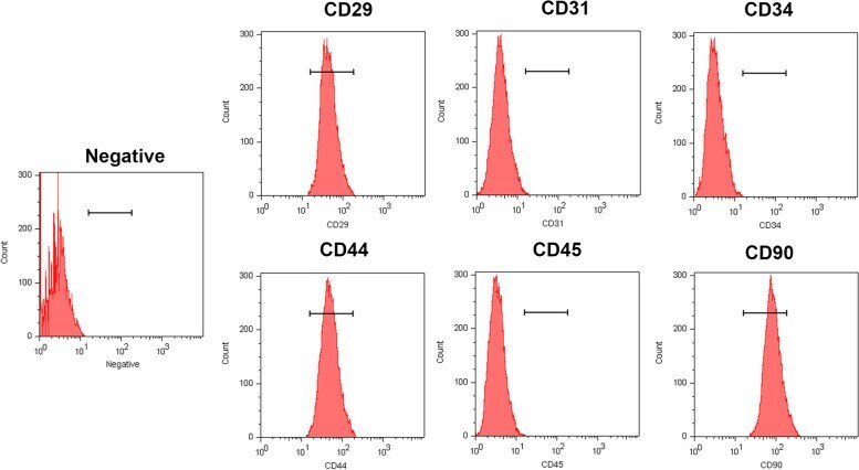
- Characterization analysis of ADSCs by flow cytometry. ADSCs were positive for mesenchymal stem cell surface antigen CD29, CD44 and CD90, but negative for hematopoietic surface antigen CD34 and CD45, and also negative for vascular endothelial cell antigen CD31
- Submitted by
- Invitrogen Antibodies (provider)
- Main image
- Experimental details
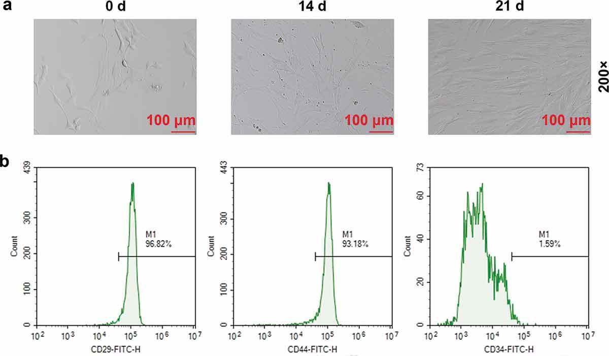
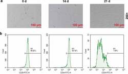
- Figure 1. Extraction and identification of bone marrow mesenchymal stem cells (BMSCs) . (a) Morphological observation of BMSCs on day 0, 14 and 21 (200x). (b) Fluorescence Activating Cell Sorter (FACS) was used to identify the contents of CD29, CD44, and CD34 in BMSCs on the 21st day