Explore
 Validate
 Learn
LearnMA5-14675
antibody from Invitrogen Antibodies
Targeting: CEACAM5
CD66e, CEA
 Western blot
 ELISA Immunocytochemistry Immunoprecipitation Flow cytometry Radioimmunoassay Other assay
Antibody data
- Antibody Data
- Antigen structure
- References [2]
- Comments [0]
- Validations
- Western blot [1]
- Immunocytochemistry [1]
- Flow cytometry [1]
- Other assay [3]
Submit
Validation data
Reference
Comment
Report error
- Product number
- MA5-14675 - Provider product page

- Provider
- Invitrogen Antibodies
- Product name
- CEA Monoclonal Antibody (1106)
- Antibody type
- Monoclonal
- Antigen
- Purifed from natural sources
- Description
- The predicted MW of CEA is ~77kD, but by Western blot MA5-14675 detects CEA with varying degrees of glycosylation at ~77-180kD. Product MA514675 is a smaller package size of MIC0102 (formerly sold as a Seradyn product).
- Reactivity
- Human
- Host
- Mouse
- Isotype
- IgG
- Antibody clone number
- 1106
- Vial size
- 100 µg
- Concentration
- 1 mg/mL
- Storage
- Maintain refrigerated at 2-8°C for up to 6 months. For long term storage store at -20°C
Submitted references Selection and characterisation of Affimers specific for CEA recognition.
Mutated CEACAMs Disrupt Transforming Growth Factor Beta Signaling and Alter the Intestinal Microbiome to Promote Colorectal Carcinogenesis.
Shamsuddin SH, Jayne DG, Tomlinson DC, McPherson MJ, Millner PA
Scientific reports 2021 Jan 12;11(1):744
Scientific reports 2021 Jan 12;11(1):744
Mutated CEACAMs Disrupt Transforming Growth Factor Beta Signaling and Alter the Intestinal Microbiome to Promote Colorectal Carcinogenesis.
Gu S, Zaidi S, Hassan MI, Mohammad T, Malta TM, Noushmehr H, Nguyen B, Crandall KA, Srivastav J, Obias V, Lin P, Nguyen BN, Yao M, Yao R, King CH, Mazumder R, Mishra B, Rao S, Mishra L
Gastroenterology 2020 Jan;158(1):238-252
Gastroenterology 2020 Jan;158(1):238-252
No comments: Submit comment
Supportive validation
- Submitted by
- Invitrogen Antibodies (provider)
- Main image
- Experimental details
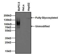
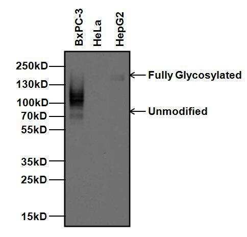
- Western blot analysis of Carcinoembryonic Antigen (CEA) was performed by loading 20 µg of BxPC-3, HeLa, and HepG2 whole cell lysates per well and 5 µL of PageRuler Plus Prestained Protein Ladder (Product # 26619) onto a 4-20% Tris-Glycine polyacrylamide gel. Proteins were transferred to a nitrocellulose membrane using the G2 Blotter (Product # 62288), and blocked with 5% Milk in TBST for 1 hour at room temperature. CEA (with varying degrees of glycosylation) was detected at ~77-180 kD using a CEA monoclonal antibody (Product # MA5-14675) at a dilution of 1 µg/mL in 5% milk in TBST overnight at 4C on a rocking platform, followed by a goat anti-mouse IgG HRP secondary antibody (Product # 31430) at a dilution of 1:10,000 for at least 30 minutes at room temperature. Chemiluminescent detection was performed using SuperSignal West Pico substrate (Product # 34080).
Supportive validation
- Submitted by
- Invitrogen Antibodies (provider)
- Main image

- Experimental details
- Immunofluorescent analysis of Carcinoembryonic Antigen (CEA, green) in BxPC-3 cells. Cells were fixed with 4% paraformaldehyde, permeabilized with 0.1% Triton X-100, and blocked with 0.3% BSA in PBS, each for 15 minutes at room temperature. Cells were stained with a CEA monoclonal antibody (Product # MA5-14675) at a dilution of 10 µg/mL in blocking buffer for 1 hour at room temperature, and then incubated with a goat anti-mouse IgG Superclonal secondary antibody, Alexa Fluor® 488 conjugate (Product # A28175) at a dilution of 1:1000 for 1 hour at room temperature. Nuclei (blue) were stained with Hoechst nuclear stain. Images were taken on a Thermo Scientific ToxInsight Instrument at 20X magnification.

Supportive validation
- Submitted by
- Invitrogen Antibodies (provider)
- Main image

- Experimental details
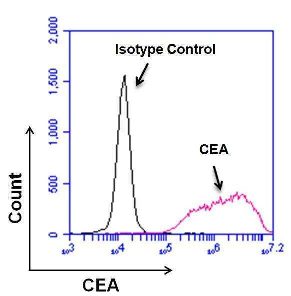
- Flow cytometry analysis of Carcinoembryonic Antigen (CEA) on BxPC-3 cells. Cells were harvested with 0.25% Trypsin-EDTA and stained with a CEA monoclonal antibody (Product # MA5-14675) at a dilution of 10 µg/mL (pink histogram), or with a mouse isotype control (black histogram) at a dilution of 10 µg/mL in PBS + 5% FCS. After incubation of the primary antibody for 1 hour on ice, the cells were stained with a goat anti-mouse IgG secondary antibody, DyLight 488 conjugate (Product # 35502) at a dilution of 1:40 for 1 hour on ice. A representative 10,000 cells were acquired for each sample.
Supportive validation
- Submitted by
- Invitrogen Antibodies (provider)
- Main image
- Experimental details
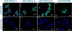
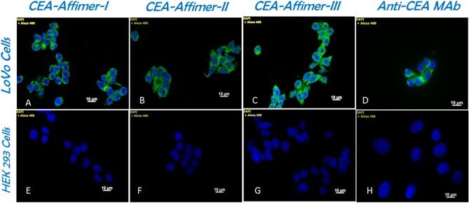
- Figure 2 Cell binding and selectivity of anti-CEA Affimers on LoVo cells in comparison to monoclonal antibody. ( A ) to ( C ) show affinity-fluorescence staining of CEA binding Affimer I, II and III on LoVo cells compared to immunofluorescence staining using anti-CEA monoclonal antibody ( D ). No binding is present on corresponding HEK 293 negative control cells ( E to H ).
- Submitted by
- Invitrogen Antibodies (provider)
- Main image

- Experimental details
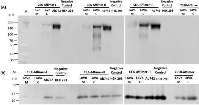
- Figure 3 Immunoblotting of Affimer precipitated CEA. Monoclonal anti-CEA and anti-His 6 tag antibodies were used to probe ( A ) the CEA protein and ( B ) Affimers, respectively from the pull-down complex. Yeast SUMO-10 binder was used as a control Affimer. Commercial CEA from Abcam was used as positive control and cell lysates from HEK 293 as negative control. LoVo M and C are samples from medium and cell lysates, respectively. Full-length blots are presented in Supplementary Fig. S3 .
- Submitted by
- Invitrogen Antibodies (provider)
- Main image

- Experimental details
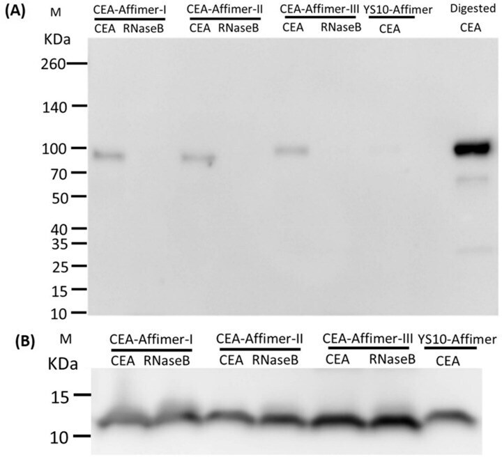
- Figure 4 Immunoblotting of Affimer precipitated deglycosylated CEA. Immunoblotting of affinity-precipitation using CEA binding Affimers as ligand to pull-down deglycosylated CEA (n = 2). Monoclonal anti-CEA and anti-His 6 tag antibodies were used to probe the pulled-down ( A ) deglycosylated CEA and ( B ) Affimers, respectively. Yeast SUMO-10 binder was used as a control Affimer and RNase B was used as non-specific analyte. Digested CEA was used as a reference to the pulled-down CEA in blotting. Full-length blot of Fig. 4B is presented in Supplementary Fig. S3 .