Explore
 Validate
 Learn
Learn Western blot
 Flow cytometry
Antibody data
- Antibody Data
- Antigen structure
- References [2]
- Comments [0]
- Validations
- Western blot [1]
- Immunocytochemistry [1]
- Immunohistochemistry [1]
Submit
Validation data
Reference
Comment
Report error
- Product number
- 14-0669-82 - Provider product page

- Provider
- Invitrogen Antibodies
- Product name
- CD66e (CEA) Monoclonal Antibody (CB30), eBioscience™
- Antibody type
- Monoclonal
- Antigen
- Other
- Description
- Description: The monoclonal antibody CB30 recognizes human CD66e, also known as CEA (carcinoembryonic antigen) and CEACAM5. As a member of the immunoglobulin superfamily, it is large GPI-linked glycosylated protein with a molecular weight about 200 kDa. It is expressed mainly on epithelial cells and on a variety of tumors of the gastrointestinal, respiratory and genitourinary tracts. CD66e has been shown to function in cell adhesion and migration via homophilic and heterophilic interactions with NCA (non-specific cross-reacting antigen also known as CEACAM6 and CD66c). Overexpression is correlated with tumor invasiveness and is therefore used as a clinical marker. Applications Reported: This CB30 antibody has been reported for use in flow cytometric analysis, immunoblotting (WB), and immunohistochemical staining. Applications Tested: This CB30 antibody has been tested by immunohistochemistry and by western blot on cell lysates prepared from MCF7 cells. This antibody can be used at less than or equal to 1 µg/mL for immunoblotting and less than 10 µg/mL for IHC. It is recommended that this antibody be carefully titrated for optimal performance in the assay of interest. Purity: Greater than 90%, as determined by SDS-PAGE. Aggregation: Less than 10%, as determined by HPLC. Filtration: 0.2 µm post-manufacturing filtered.
- Reactivity
- Human
- Host
- Mouse
- Isotype
- IgG
- Antibody clone number
- CB30
- Vial size
- 100 µg
- Concentration
- 0.5 mg/mL
- Storage
- 4° C
Submitted references Carcinoembryonic antigen gene family: molecular biology and clinical perspectives.
Carcinoembryonic antigen, a human tumor marker, functions as an intercellular adhesion molecule.
Thompson JA, Grunert F, Zimmermann W
Journal of clinical laboratory analysis 1991;5(5):344-66
Journal of clinical laboratory analysis 1991;5(5):344-66
Carcinoembryonic antigen, a human tumor marker, functions as an intercellular adhesion molecule.
Benchimol S, Fuks A, Jothy S, Beauchemin N, Shirota K, Stanners CP
Cell 1989 Apr 21;57(2):327-34
Cell 1989 Apr 21;57(2):327-34
No comments: Submit comment
Supportive validation
- Submitted by
- Invitrogen Antibodies (provider)
- Main image
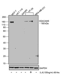
- Experimental details
- Western blot was performed using Anti-CD66e (CEA) Monoclonal Antibody (CB30), eBioscience™ (Product # 14-0669-82) and a 160 kDa band corresponding to Carcinoembryonic antigen-related cell adhesion molecule 5 was observed across cell lines tested. Membrane enriched extracts (30 µg lysate) of MCF7 (Lane 1), HCT 116 (Lane 2), SW480 (Lane 3), HT-29 (Lane 4), HT-29 treated with IL-6, 100 ng/mL, 48 Hrs. (Lane 5), MDA-MB-231 (Lane 6) were electrophoresed using NuPAGE™ 4-12% Bis-Tris Protein Gel (Product # NP0322BOX). Resolved proteins were then transferred onto a Nitrocellulose membrane (Product # IB23001) by iBlot® 2 Dry Blotting System (Product # IB21001). The blot was probed with the primary antibody (1:1000 Dilution) and detected by chemiluminescence with Goat anti-Mouse IgG (H+L) Superclonal™ Recombinant Secondary Antibody, HRP (Product # A28177, 1:4000 dilution) using the iBright FL 1000 (Product # A32752). Chemiluminescent detection was performed using Novex® ECL Chemiluminescent Substrate Reagent Kit (Product # WP20005).

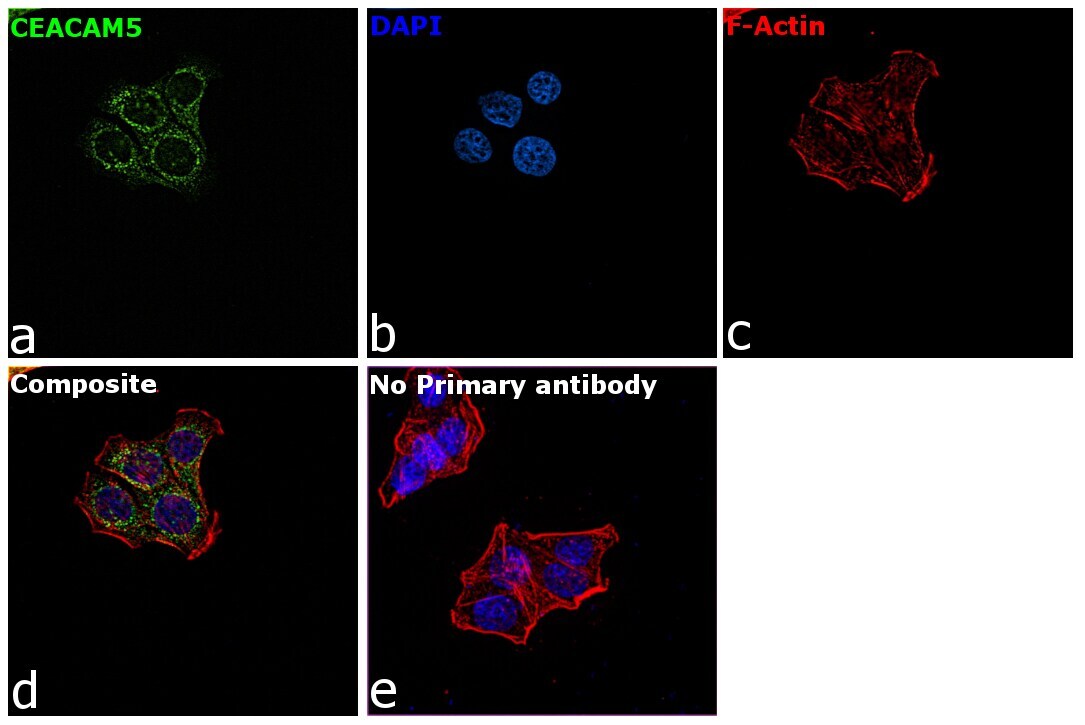
Supportive validation
- Submitted by
- Invitrogen Antibodies (provider)
- Main image
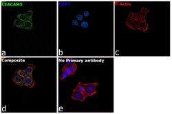
- Experimental details
- Immunofluorescence analysis of Carcinoembryonic antigen-related cell adhesion molecule 5 was performed using 70% confluent log phase HT-29 cells. The cells were fixed with 4% paraformaldehyde for 10 minutes, permeabilized with 0.1% Triton™ X-100 for 15 minutes, and blocked with 2% BSA for 45 minutes at room temperature. The cells were labeled with CD66e (CEA) Monoclonal Antibody (CB30), eBioscience™ (Product # 14-0669-82) at 5 µg/mL in 0.1% BSA, incubated at 4 degree celsius overnight and then labeled with Goat anti-Mouse IgG (H+L) Superclonal™ Recombinant Secondary Antibody, Alexa Fluor® 488 conjugate (Product # A28175), (1:2000 dilution), for 45 minutes at room temperature (Panel a: Green). Nuclei (Panel b:Blue) were stained with ProLong™ Diamond Antifade Mountant with DAPI (Product # P36962). F-actin (Panel c: Red) was stained with Rhodamine Phalloidin (Product # R415, 1:300 dilution). Panel d represents the merged image showing cell membrane localization. Panel e represents control cells with no primary antibody to assess background. The images were captured at 60x magnification.
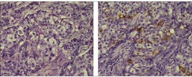
Supportive validation
- Submitted by
- Invitrogen Antibodies (provider)
- Main image
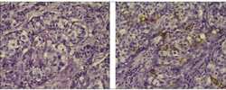
- Experimental details
- Immunohistochemistry of formalin-fixed paraffin embedded human infiltrating ductal carcinoma, using 10 µg/mL of Mouse IgG1 Isotype Control Purified (Product # 14-4714-82) (left) or Anti-Human CD66 (CEA) Purified (right) followed by Anti-Mouse Biotin, and DAB visualization. Nuclei are counterstained with hematoxylin.