 Explore
 Validate
 Learn
LearnPA5-16665
antibody from Invitrogen Antibodies
Targeting: CEACAM5
CD66e, CEA
 Western blot
 Immunocytochemistry Immunoprecipitation Immunohistochemistry Flow cytometry Other assay
Antibody data
- Antibody Data
- Antigen structure
- References [3]
- Comments [0]
- Validations
- Immunocytochemistry [1]
- Immunohistochemistry [1]
- Flow cytometry [3]
- Other assay [2]
Submit
Validation data
Reference
Comment
Report error
- Product number
- PA5-16665 - Provider product page

- Provider
- Invitrogen Antibodies
- Product name
- Anti-CEA Polyclonal Antibody
- Antibody type
- Polyclonal
- Antigen
- Other
- Description
- PA5-16668 targets Lysozyme in IHC (P) applications and shows reactivity with Canine, Feline, Human, Non-human primate, and Porcine samples. The PA5-16668 immunogen is human lysozyme purified from the urine of patients with monocytic leukemia.
- Reactivity
- Human, Mouse
- Host
- Rabbit
- Isotype
- IgG
- Vial size
- 500 µL
- Concentration
- Lot Dependent
- Storage
- 4° C
Submitted references Establishment of three novel cell lines derived from African American patients with colorectal carcinoma: A unique tool for assessing racial health disparity.
New diagnostic markers in salivary gland tumors.
Prognostic and predictive markers in medullary thyroid carcinoma.
Paredes J, Ji P, Lacomb JF, Shroyer KR, Martello LA, Williams JL
International journal of oncology 2018 Oct;53(4):1516-1528
International journal of oncology 2018 Oct;53(4):1516-1528
New diagnostic markers in salivary gland tumors.
Schneider S, Kloimstein P, Pammer J, Brannath W, Grasl MCh, Erovic BM
European archives of oto-rhino-laryngology : official journal of the European Federation of Oto-Rhino-Laryngological Societies (EUFOS) : affiliated with the German Society for Oto-Rhino-Laryngology - Head and Neck Surgery 2014 Jul;271(7):1999-2007
European archives of oto-rhino-laryngology : official journal of the European Federation of Oto-Rhino-Laryngological Societies (EUFOS) : affiliated with the German Society for Oto-Rhino-Laryngology - Head and Neck Surgery 2014 Jul;271(7):1999-2007
Prognostic and predictive markers in medullary thyroid carcinoma.
Erovic BM, Kim D, Cassol C, Goldstein DP, Irish JC, Asa SL, Mete O
Endocrine pathology 2012 Dec;23(4):232-42
Endocrine pathology 2012 Dec;23(4):232-42
No comments: Submit comment
Supportive validation
- Submitted by
- Invitrogen Antibodies (provider)
- Main image

- Experimental details
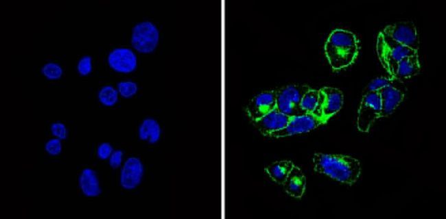
- Immunofluorescent analysis of CD66e (green) showing staining in the cytoplasm and membrane of HepG2 cells (right) compared to a negative control without primary antibody (left). Formalin-fixed cells were permeabilized with 0.1% Triton X-100 in TBS for 5-10 minutes and blocked with 3% BSA-PBS for 30 minutes at room temperature. Cells were probed with a CD66e polyclonal antibody (Product # PA5-16665) in 3% BSA-PBS at a dilution of 1:100 and incubated overnight at 4 ºC in a humidified chamber. Cells were washed with PBST and incubated with a DyLight-conjugated secondary antibody in PBS at room temperature in the dark. F-actin (red) was stained with a flourescent red phalloidin and nuclei (blue) were stained with Hoechst or DAPI. Images were taken at a magnification of 60x.
Supportive validation
- Submitted by
- Invitrogen Antibodies (provider)
- Main image
- Experimental details
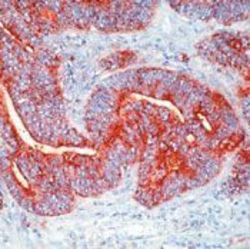
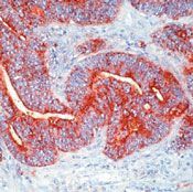
- Formalin-fixed, paraffin-embedded human colon carcinoma stained with Carcinoembryonic Antigen antibody using peroxidase-conjugate and AEC chromogen. Note luminal surface and cytoplasmic staining of tumor cells.
Supportive validation
- Submitted by
- Invitrogen Antibodies (provider)
- Main image
- Experimental details
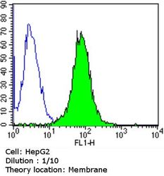
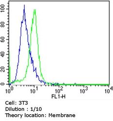
- Flow cytometry analysis of CD66e in HepG2 cells compared to an isotype control (blue). Cells were harvested, adjusted to a concentration of 1-5x10^6 cells/ml, fixed with 2% paraformaldehyde and washed with PBS. Cells were blocked with a 2% solution of BSA-PBS for 30 min at room temperature and incubated with a CD66e Polyclonal Antibody (Product # PA5-16665) at a dilution of 1:10 for 60 min at room temperature. Cells were then incubated for 40 min at room temperature in the dark using a Dylight 488-conjugated goat anti-rabbit IgG (H+L) secondary antibody and re-suspended in PBS for FACS analysis.

- Submitted by
- Invitrogen Antibodies (provider)
- Main image
- Experimental details
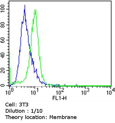
- Flow cytometry analysis of CD66e in NIH/3T3 cells compared to an isotype control (blue). Cells were harvested, adjusted to a concentration of 1-5x10^6 cells/ml, fixed with 2% paraformaldehyde and washed with PBS. Cells were blocked with a 2% solution of BSA-PBS for 30 min at room temperature and incubated with a CD66e Polyclonal Antibody (Product # PA5-16665) at a dilution of 1:10 for 60 min at room temperature. Cells were then incubated for 40 min at room temperature in the dark using a Dylight 488-conjugated goat anti-rabbit IgG (H+L) secondary antibody and re-suspended in PBS for FACS analysis.
- Submitted by
- Invitrogen Antibodies (provider)
- Main image

- Experimental details
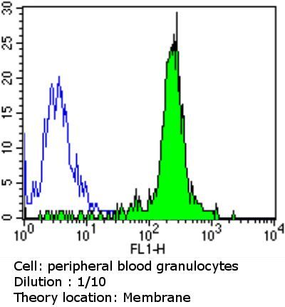
- Flow cytometry analysis of CD66e in blood granulocytes compared to an isotype control (blue). Human blood was collected, combined with a hydrophilic polysaccharide, centrifuged, transferred to a conical tube and washed with PBS. 50 ul of cell solution was added to each tube at a dilution of 2x10^7 cells/ml, followed by the addition of 50 ul of isotype control and primary antibody (Product # PA5-16665) at a dilution of 1:10. Cells were incubated for 30 min at 4ºC and washed with a cell buffer, followed by incubation with a DyLight 488-conjugated goat anti-rabbit IgG (H+L) secondary for 30 min at 4ºC in the dark. FACS analysis was performed using 400 ul of cell buffer.
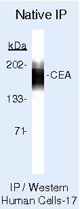
Supportive validation
- Submitted by
- Invitrogen Antibodies (provider)
- Main image
- Experimental details
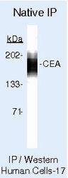
- Immunoprecipitation of CD66e using CD66e Polyclonal Antibody (Product # PA5-16665) on Native Human LS174T Cells.
- Submitted by
- Invitrogen Antibodies (provider)
- Main image
- Experimental details
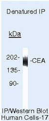
- Immunoprecipitation of CD66e using CD66e Polyclonal Antibody (Product # PA5-16665) on denatured Human LS174T Cells.
