Explore
 Validate
 Learn
Learn Western blot
 Immunocytochemistry
Antibody data
- Antibody Data
- Antigen structure
- References [1]
- Comments [0]
- Validations
- Immunocytochemistry [2]
- Immunohistochemistry [1]
- Other assay [2]
Submit
Validation data
Reference
Comment
Report error
- Product number
- PA5-15275 - Provider product page

- Provider
- Invitrogen Antibodies
- Product name
- PI4K2A Polyclonal Antibody
- Antibody type
- Polyclonal
- Antigen
- Synthetic peptide
- Reactivity
- Human, Mouse
- Host
- Rabbit
- Isotype
- IgG
- Vial size
- 400 μL
- Concentration
- 1 mg/mL
- Storage
- Store at 4°C short term. For long term storage, store at -20°C, avoiding freeze/thaw cycles.
Submitted references Coupling of microtubule motors with AP-3 generated organelles in axons by NEEP21 family member calcyon.
Shi L, Hines T, Bergson C, Smith D
Molecular biology of the cell 2018 Aug 15;29(17):2055-2068
Molecular biology of the cell 2018 Aug 15;29(17):2055-2068
No comments: Submit comment
Supportive validation
- Submitted by
- Invitrogen Antibodies (provider)
- Main image
- Experimental details
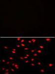
- Immunofluorescent analysis using a PI4K2A polyclonal antibody (Product # PA5-15275) on HeLa cells. 0.025 mg/mL primary antibody was followed by fluor-conjugated donkey anti-rabbit lgG (H+L).

- Submitted by
- Invitrogen Antibodies (provider)
- Main image

- Experimental details
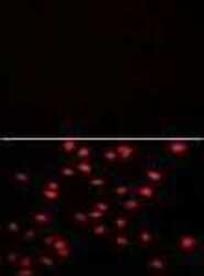
- Immunocytochemistry analysis of PI4K2A in HeLa cells. Samples were incubated with PI4K2A polyclonal antibody (Product # PA5-15275) using a dilution of 0.025 mg/mL followed by Alexa-Fluor-546-conjugated donkey anti-rabbit lgG (H+L). Negative control of Hela cells without Alexa-Fluor-546-conjugated donkey anti-rabbit lgG (H+L). Alexa-Fluor-546 emits orange fluorescence.
Supportive validation
- Submitted by
- Invitrogen Antibodies (provider)
- Main image

- Experimental details
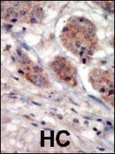
- Immunohistochemistry analysis of PI4K2A in formalin-fixed and paraffin-embedded human cancer tissue. Samples were incubated with PI4K2A polyclonal antibody (Product # PA5-15275) which was peroxidase-conjugated to the secondary antibody, followed by DAB staining. This data demonstrates the use of this antibody for immunohistochemistry; clinical relevance has not been evaluated. BC = breast carcinoma; HC = hepatocarcinoma.
Supportive validation
- Submitted by
- Invitrogen Antibodies (provider)
- Main image
- Experimental details
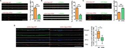
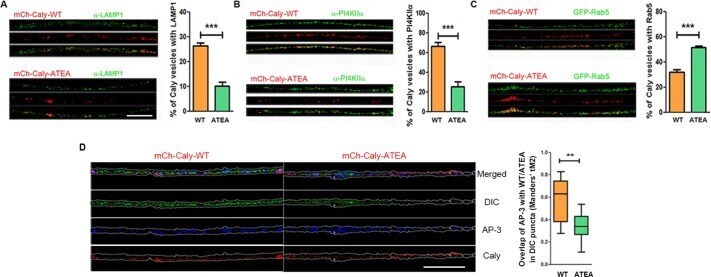
- FIGURE 4: Caly sorting requires interaction with adaptor protein complexes. (A-C) Differential localization of Caly-WT and Caly-ATEA in EE/SEs and LE/LROs. Confocal micrographs of DRG axons transfected with mCh-Caly-WT or mCh-Caly-ATEA (red) and stained with LAMP1 (A) or PI4KIIalpha (B) antibodies or cotransfected with GFP-Rab5 (C) (green). Bar graphs show the mean and SEM of overlapping red and green puncta in 100-mum axon segments of each group. (D) Confocal micrographs of DRG axons transfected with mCh-Caly-WT or mCh-Caly-ATEA (red) and stained with DIC (green) and AP-3 (blue). Manders coefficient of overlap was determined for colocalization of Caly-WT or Caly-ATEA with AP-3 in DIC positive puncta. Caly-WT exhibited greater colocalization with AP-3/DIC positive puncta than Caly-ATEA. Box-and-whisker plots show the Manders's tM2 of overlapping red and blue puncta in 100-mum axon segments of each group. Data plotted in histograms or in box-and-whisker plots correspond to results obtained in three independent experiments from at least five axons per experiment for each group; ** p < 0.01, *** p < 0.001; bar = 20 mum.
- Submitted by
- Invitrogen Antibodies (provider)
- Main image
- Experimental details
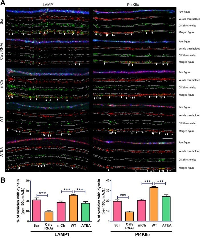
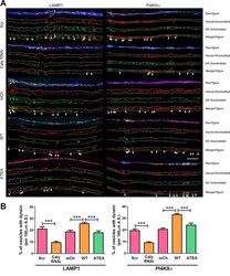
- FIGURE 5: Caly stimulated dynein recruitment requires interaction with adaptor protein complexes. (A) Dynein labeling of LE/LROs in DRG axons transfected with GFP-Caly RNAi, GFP-Scr RNAi, mCherry vector, mCh-Caly-WT, or mCh-Caly-ATEA and stained with LAMP1 and PI4KIIalpha antibodies. Axons are outlined in white; filled arrows point to overlapping puncta, whereas hollow arrows show the examples of nonoverlapping puncta. (B) Greater dynein colocalization with LAMP1- or PI4KIIalpha-positive organelles is detected in Caly-WT-transfected axons, whereas less is observed in Caly RNAi-transfected axons. Data plotted in bar graphs correspond to results obtained in three independent experiments from at least five axons per experiment for each group; *** p < 0.001; bar = 10 mum.