Explore
 Validate
 Learn
Learn Western blot
 Immunocytochemistry
Antibody data
- Antibody Data
- Antigen structure
- References [42]
- Comments [0]
- Validations
- Immunocytochemistry [4]
- Immunohistochemistry [3]
- Flow cytometry [1]
- Other assay [10]
Submit
Validation data
Reference
Comment
Report error
- Product number
- MA5-12596 - Provider product page

- Provider
- Invitrogen Antibodies
- Product name
- Cytokeratin 5 Monoclonal Antibody (XM26)
- Antibody type
- Monoclonal
- Antigen
- Recombinant full-length protein
- Description
- MA5-12596 targets Cytokeratin 5 in IHC (P) applications and shows reactivity with Human samples. The MA5-12596 immunogen is recombinant protein corresponding to C-terminal 103 aa of cytokeratin 5.
- Reactivity
- Human
- Host
- Mouse
- Isotype
- IgG
- Antibody clone number
- XM26
- Vial size
- 500 μL
- Concentration
- Conc. Not Determined
- Storage
- 4°C
Submitted references Fibulin-4 Accelerates Amyloid Formation by Binding with a Keratin 5 Peptide Fragment.
Common tumor-suppressive signaling of thyroid hormone receptor beta in breast and thyroid cancer cells.
High Ki-67 expression is a marker of poor survival in apocrine breast carcinoma.
Long-Term Modeling of SARS-CoV-2 Infection of In Vitro Cultured Polarized Human Airway Epithelium.
Long Period Modeling SARS-CoV-2 Infection of in Vitro Cultured Polarized Human Airway Epithelium.
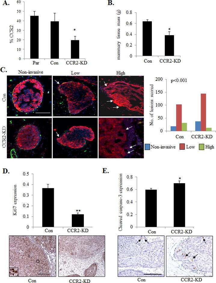
CCR2 Chemokine Receptors Enhance Growth and Cell-Cycle Progression of Breast Cancer Cells through SRC and PKC Activation.
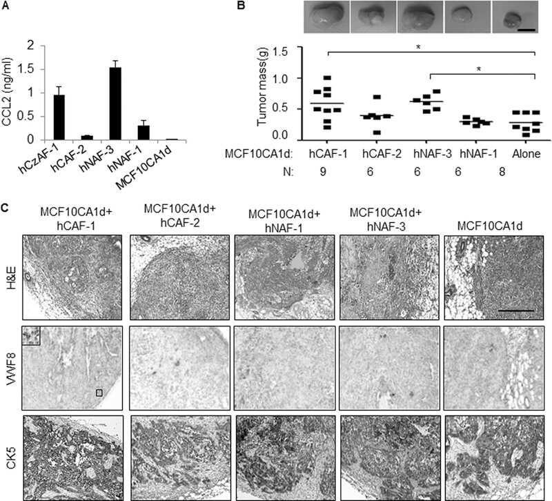
Chemokine Signaling Facilitates Early-Stage Breast Cancer Survival and Invasion through Fibroblast-Dependent Mechanisms.
Continuous Delivery of Neutralizing Antibodies Elevate CCL2 Levels in Mice Bearing MCF10CA1d Breast Tumor Xenografts.
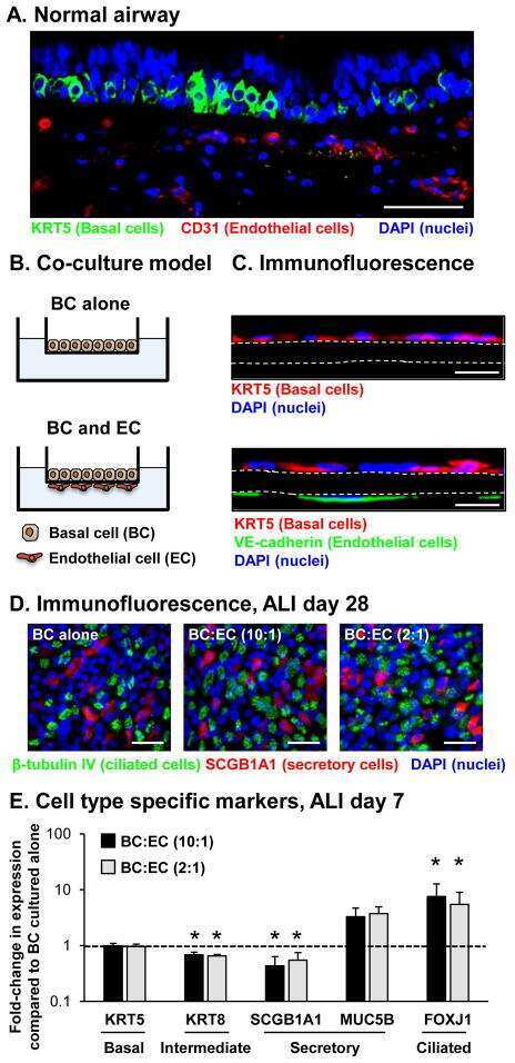
Endothelial Cell Mediated Promotion of Ciliated Cell Differentiation of Human Airway Basal Cells via Insulin and Insulin-Like Growth Factor 1 Receptor Mediated Signaling.
FAM83H and casein kinase I regulate the organization of the keratin cytoskeleton and formation of desmosomes.
Steroid induction of therapy-resistant cytokeratin-5-positive cells in estrogen receptor-positive breast cancer through a BCL6-dependent mechanism.
Targeted Pten deletion plus p53-R270H mutation in mouse mammary epithelium induces aggressive claudin-low and basal-like breast cancer.
High frequency of loss of PTEN expression in human solid salivary adenoid cystic carcinoma and its implication for targeted therapy.
Biological characteristics and clinical outcome of triple negative primary breast cancer in older women - comparison with their younger counterparts.
A systematic analysis of commonly used antibodies in cancer diagnostics.
HIV nucleoside reverse transcriptase inhibitors efavirenz and tenofovir change the growth and differentiation of primary gingival epithelium.
Prolactin suppresses a progestin-induced CK5-positive cell population in luminal breast cancer through inhibition of progestin-driven BCL6 expression.
Immunohistochemical profile of high-grade ductal carcinoma in situ of the breast.
Genomic and phenotypic profiles of two Brazilian breast cancer cell lines derived from primary human tumors.
Tumor grade and matrix metalloproteinase 2 expression in stromal fibroblasts help to stratify the high-risk group of patients with early breast cancer identified on the basis of st Gallen recommendations.
Effect of the HIV nucleoside reverse transcriptase inhibitor zidovudine on the growth and differentiation of primary gingival epithelium.
Two novel recessive mutations in KRT14 identified in a cohort of 21 Spanish families with epidermolysis bullosa simplex.
Molecular phenotypes of matched in situ and invasive components of breast carcinomas.
Identification of the receptor tyrosine kinase AXL in breast cancer as a target for the human miR-34a microRNA.
Cell adhesion and communication proteins are differentially expressed in melanoma progression model.
The HIV protease inhibitor lopinavir/ritonavir (Kaletra) alters the growth, differentiation and proliferation of primary gingival epithelium.
Tumor-in-tumor of the thyroid with basaloid differentiation: a lesion with a solid cell nest neoplastic component?
Increased NOS2 predicts poor survival in estrogen receptor-negative breast cancer patients.
Molecular characterization of breast cancer in young Brazilian women.
Molecular characterization of breast cancer in young Brazilian women.
Differences in the tumor microenvironment between African-American and European-American breast cancer patients.
Expression and prognostic significance of metalloproteases and their inhibitors in luminal A and basal-like phenotypes of breast carcinoma.
Vessel density assessed by endoglin expression in breast carcinomas with different expression profiles.
Cytological criteria to predict basal phenotype of breast carcinomas.
Basal phenotype in breast carcinoma occurring in women aged 35 or younger.
Keratin profiling in the developing human prostate. A different approach to understanding epithelial lineage.
Identification of molecular phenotypes in canine mammary carcinomas with clinical implications: application of the human classification.
Tissue microarrays for testing basal biomarkers in familial breast cancer cases.
P-cadherin and cytokeratin 5: useful adjunct markers to distinguish basal-like ductal carcinomas in situ.
Nestin is expressed in the basal/myoepithelial layer of the mammary gland and is a selective marker of basal epithelial breast tumors.
Adenosquamous carcinoma with cilium formation, mucin production and keratinization in the nasal cavity of a red fox (Vulpes vulpes schrencki).
p63, cytokeratin 5, and P-cadherin: three molecular markers to distinguish basal phenotype in breast carcinomas.
Katagiri F, Ueo D, Okubo-Gunge Y, Usui A, Kuwatsuka S, Mine Y, Hamada K, Fujiwara S, Sasaki T, Nomizu M, Utani A
JID innovations : skin science from molecules to population health 2022 May;2(3):100114
JID innovations : skin science from molecules to population health 2022 May;2(3):100114
Common tumor-suppressive signaling of thyroid hormone receptor beta in breast and thyroid cancer cells.
Bolf EL, Gillis NE, Davidson CD, Cozzens LM, Kogut S, Tomczak JA, Frietze S, Carr FE
Molecular carcinogenesis 2021 Dec;60(12):874-885
Molecular carcinogenesis 2021 Dec;60(12):874-885
High Ki-67 expression is a marker of poor survival in apocrine breast carcinoma.
Wysocka J, Adamczyk A, Kruczak A, Niemiec J, Sas-Korczyńska B
Polish journal of pathology : official journal of the Polish Society of Pathologists 2020;71(2):107-119
Polish journal of pathology : official journal of the Polish Society of Pathologists 2020;71(2):107-119
Long-Term Modeling of SARS-CoV-2 Infection of In Vitro Cultured Polarized Human Airway Epithelium.
Hao S, Ning K, Kuz CA, Vorhies K, Yan Z, Qiu J
mBio 2020 Nov 6;11(6)
mBio 2020 Nov 6;11(6)
Long Period Modeling SARS-CoV-2 Infection of in Vitro Cultured Polarized Human Airway Epithelium.
Hao S, Ning K, Kuz CA, Vorhies K, Yan Z, Qiu J
bioRxiv : the preprint server for biology 2020 Aug 28;
bioRxiv : the preprint server for biology 2020 Aug 28;
CCR2 Chemokine Receptors Enhance Growth and Cell-Cycle Progression of Breast Cancer Cells through SRC and PKC Activation.
Yao M, Fang W, Smart C, Hu Q, Huang S, Alvarez N, Fields P, Cheng N
Molecular cancer research : MCR 2019 Feb;17(2):604-617
Molecular cancer research : MCR 2019 Feb;17(2):604-617
Chemokine Signaling Facilitates Early-Stage Breast Cancer Survival and Invasion through Fibroblast-Dependent Mechanisms.
Brummer G, Acevedo DS, Hu Q, Portsche M, Fang WB, Yao M, Zinda B, Myers M, Alvarez N, Fields P, Hong Y, Behbod F, Cheng N
Molecular cancer research : MCR 2018 Feb;16(2):296-308
Molecular cancer research : MCR 2018 Feb;16(2):296-308
Continuous Delivery of Neutralizing Antibodies Elevate CCL2 Levels in Mice Bearing MCF10CA1d Breast Tumor Xenografts.
Yao M, Smart C, Hu Q, Cheng N
Translational oncology 2017 Oct;10(5):734-743
Translational oncology 2017 Oct;10(5):734-743
Endothelial Cell Mediated Promotion of Ciliated Cell Differentiation of Human Airway Basal Cells via Insulin and Insulin-Like Growth Factor 1 Receptor Mediated Signaling.
Gomi K, Tang Y, Arbelaez V, Crystal RG, Walters MS
Stem cell reviews and reports 2017 Apr;13(2):309-317
Stem cell reviews and reports 2017 Apr;13(2):309-317
FAM83H and casein kinase I regulate the organization of the keratin cytoskeleton and formation of desmosomes.
Kuga T, Sasaki M, Mikami T, Miake Y, Adachi J, Shimizu M, Saito Y, Koura M, Takeda Y, Matsuda J, Tomonaga T, Nakayama Y
Scientific reports 2016 May 25;6:26557
Scientific reports 2016 May 25;6:26557
Steroid induction of therapy-resistant cytokeratin-5-positive cells in estrogen receptor-positive breast cancer through a BCL6-dependent mechanism.
Goodman CR, Sato T, Peck AR, Girondo MA, Yang N, Liu C, Yanac AF, Kovatich AJ, Hooke JA, Shriver CD, Mitchell EP, Hyslop T, Rui H
Oncogene 2016 Mar 17;35(11):1373-85
Oncogene 2016 Mar 17;35(11):1373-85
Targeted Pten deletion plus p53-R270H mutation in mouse mammary epithelium induces aggressive claudin-low and basal-like breast cancer.
Wang S, Liu JC, Kim D, Datti A, Zacksenhaus E
Breast cancer research : BCR 2016 Jan 19;18(1):9
Breast cancer research : BCR 2016 Jan 19;18(1):9
High frequency of loss of PTEN expression in human solid salivary adenoid cystic carcinoma and its implication for targeted therapy.
Liu H, Du L, Wang R, Wei C, Liu B, Zhu L, Liu P, Liu Q, Li J, Lu SL, Xiao J
Oncotarget 2015 May 10;6(13):11477-91
Oncotarget 2015 May 10;6(13):11477-91
Biological characteristics and clinical outcome of triple negative primary breast cancer in older women - comparison with their younger counterparts.
Syed BM, Green AR, Nolan CC, Morgan DA, Ellis IO, Cheung KL
PloS one 2014;9(7):e100573
PloS one 2014;9(7):e100573
A systematic analysis of commonly used antibodies in cancer diagnostics.
Gremel G, Bergman J, Djureinovic D, Edqvist PH, Maindad V, Bharambe BM, Khan WA, Navani S, Elebro J, Jirström K, Hellberg D, Uhlén M, Micke P, Pontén F
Histopathology 2014 Jan;64(2):293-305
Histopathology 2014 Jan;64(2):293-305
HIV nucleoside reverse transcriptase inhibitors efavirenz and tenofovir change the growth and differentiation of primary gingival epithelium.
Mitchell D, Israr M, Alam S, Dinello D, Kishel J, Jia R, Meyers C
HIV medicine 2014 Apr;15(4):196-202
HIV medicine 2014 Apr;15(4):196-202
Prolactin suppresses a progestin-induced CK5-positive cell population in luminal breast cancer through inhibition of progestin-driven BCL6 expression.
Sato T, Tran TH, Peck AR, Girondo MA, Liu C, Goodman CR, Neilson LM, Freydin B, Chervoneva I, Hyslop T, Kovatich AJ, Hooke JA, Shriver CD, Fuchs SY, Rui H
Oncogene 2014 Apr 24;33(17):2215-24
Oncogene 2014 Apr 24;33(17):2215-24
Immunohistochemical profile of high-grade ductal carcinoma in situ of the breast.
Perez AA, Rocha RM, Balabram D, Souza Áda S, Gobbi H
Clinics (Sao Paulo, Brazil) 2013 May;68(5):674-8
Clinics (Sao Paulo, Brazil) 2013 May;68(5):674-8
Genomic and phenotypic profiles of two Brazilian breast cancer cell lines derived from primary human tumors.
Corrêa NC, Kuasne H, Faria JA, Seixas CC, Santos IG, Abreu FB, Nonogaki S, Rocha RM, Aparecida Borges Silva G, Gobbi H, Rogatto SR, Goes AM, Gomes DA
Oncology reports 2013 Apr;29(4):1299-307
Oncology reports 2013 Apr;29(4):1299-307
Tumor grade and matrix metalloproteinase 2 expression in stromal fibroblasts help to stratify the high-risk group of patients with early breast cancer identified on the basis of st Gallen recommendations.
Niemiec J, Adamczyk A, Małecki K, Ambicka A, Ryś J
Clinical breast cancer 2013 Apr;13(2):119-28
Clinical breast cancer 2013 Apr;13(2):119-28
Effect of the HIV nucleoside reverse transcriptase inhibitor zidovudine on the growth and differentiation of primary gingival epithelium.
Mitchell D, Israr M, Alam S, Kishel J, Dinello D, Meyers C
HIV medicine 2012 May;13(5):276-90
HIV medicine 2012 May;13(5):276-90
Two novel recessive mutations in KRT14 identified in a cohort of 21 Spanish families with epidermolysis bullosa simplex.
García M, Santiago JL, Terrón A, Hernández-Martín A, Vicente A, Fortuny C, De Lucas R, López JC, Cuadrado-Corrales N, Holguín A, Illera N, Duarte B, Sánchez-Jimeno C, Llames S, García E, Ayuso C, Martínez-Santamaría L, Castiglia D, De Luca N, Torrelo A, Mechan D, Baty D, Zambruno G, Escámez MJ, Del Río M
The British journal of dermatology 2011 Sep;165(3):683-92
The British journal of dermatology 2011 Sep;165(3):683-92
Molecular phenotypes of matched in situ and invasive components of breast carcinomas.
Martins D, Sousa B, Lopes N, Gomes M, Veronese L, Albergaria A, Paredes J, Schmitt F
Human pathology 2011 Oct;42(10):1438-46
Human pathology 2011 Oct;42(10):1438-46
Identification of the receptor tyrosine kinase AXL in breast cancer as a target for the human miR-34a microRNA.
Mackiewicz M, Huppi K, Pitt JJ, Dorsey TH, Ambs S, Caplen NJ
Breast cancer research and treatment 2011 Nov;130(2):663-79
Breast cancer research and treatment 2011 Nov;130(2):663-79
Cell adhesion and communication proteins are differentially expressed in melanoma progression model.
Rezze GG, Fregnani JH, Duprat J, Landman G
Human pathology 2011 Mar;42(3):409-18
Human pathology 2011 Mar;42(3):409-18
The HIV protease inhibitor lopinavir/ritonavir (Kaletra) alters the growth, differentiation and proliferation of primary gingival epithelium.
Israr M, Mitchell D, Alam S, Dinello D, Kishel JJ, Meyers C
HIV medicine 2011 Mar;12(3):145-56
HIV medicine 2011 Mar;12(3):145-56
Tumor-in-tumor of the thyroid with basaloid differentiation: a lesion with a solid cell nest neoplastic component?
Eloy C, Vinagre J, Cameselle-Teijeiro J, Paiva ME, Soares P, Sobrinho-Simões M
International journal of surgical pathology 2011 Apr;19(2):276-80
International journal of surgical pathology 2011 Apr;19(2):276-80
Increased NOS2 predicts poor survival in estrogen receptor-negative breast cancer patients.
Glynn SA, Boersma BJ, Dorsey TH, Yi M, Yfantis HG, Ridnour LA, Martin DN, Switzer CH, Hudson RS, Wink DA, Lee DH, Stephens RM, Ambs S
The Journal of clinical investigation 2010 Nov;120(11):3843-54
The Journal of clinical investigation 2010 Nov;120(11):3843-54
Molecular characterization of breast cancer in young Brazilian women.
Carvalho LV, Pereira EM, Frappart L, Boniol M, Bernardo WM, Tarricone V, Tavtigian S, Southey MC
Revista da Associacao Medica Brasileira (1992) 2010 May-Jun;56(3):278-87
Revista da Associacao Medica Brasileira (1992) 2010 May-Jun;56(3):278-87
Differences in the tumor microenvironment between African-American and European-American breast cancer patients.
Martin DN, Boersma BJ, Yi M, Reimers M, Howe TM, Yfantis HG, Tsai YC, Williams EH, Lee DH, Stephens RM, Weissman AM, Ambs S
PloS one 2009;4(2):e4531
PloS one 2009;4(2):e4531
Expression and prognostic significance of metalloproteases and their inhibitors in luminal A and basal-like phenotypes of breast carcinoma.
González LO, Corte MD, Junquera S, González-Fernández R, del Casar JM, García C, Andicoechea A, Vázquez J, Pérez-Fernández R, Vizoso FJ
Human pathology 2009 Sep;40(9):1224-33
Human pathology 2009 Sep;40(9):1224-33
Vessel density assessed by endoglin expression in breast carcinomas with different expression profiles.
Lopes N, Sousa B, Vieira D, Milanezi F, Schmitt F
Histopathology 2009 Nov;55(5):594-9
Histopathology 2009 Nov;55(5):594-9
Cytological criteria to predict basal phenotype of breast carcinomas.
Dufloth RM, Alves JM, Martins D, Vieira DS, Chikota H, Zeferino LC, Schmitt F
Diagnostic cytopathology 2009 Nov;37(11):809-14
Diagnostic cytopathology 2009 Nov;37(11):809-14
Basal phenotype in breast carcinoma occurring in women aged 35 or younger.
Bori R, Cserni G
Pathology oncology research : POR 2009 Mar;15(1):41-5
Pathology oncology research : POR 2009 Mar;15(1):41-5
Keratin profiling in the developing human prostate. A different approach to understanding epithelial lineage.
Trompetter M, Smedts F, van der Wijk J, Schoots C, de Jong HJ, Hopman A, de la Rosette J
Anticancer research 2008 Jan-Feb;28(1A):237-43
Anticancer research 2008 Jan-Feb;28(1A):237-43
Identification of molecular phenotypes in canine mammary carcinomas with clinical implications: application of the human classification.
Gama A, Alves A, Schmitt F
Virchows Archiv : an international journal of pathology 2008 Aug;453(2):123-32
Virchows Archiv : an international journal of pathology 2008 Aug;453(2):123-32
Tissue microarrays for testing basal biomarkers in familial breast cancer cases.
Dufloth RM, Matos I, Schmitt F, Zeferino LC
Sao Paulo medical journal = Revista paulista de medicina 2007 Jul 5;125(4):226-30
Sao Paulo medical journal = Revista paulista de medicina 2007 Jul 5;125(4):226-30
P-cadherin and cytokeratin 5: useful adjunct markers to distinguish basal-like ductal carcinomas in situ.
Paredes J, Lopes N, Milanezi F, Schmitt FC
Virchows Archiv : an international journal of pathology 2007 Jan;450(1):73-80
Virchows Archiv : an international journal of pathology 2007 Jan;450(1):73-80
Nestin is expressed in the basal/myoepithelial layer of the mammary gland and is a selective marker of basal epithelial breast tumors.
Li H, Cherukuri P, Li N, Cowling V, Spinella M, Cole M, Godwin AK, Wells W, DiRenzo J
Cancer research 2007 Jan 15;67(2):501-10
Cancer research 2007 Jan 15;67(2):501-10
Adenosquamous carcinoma with cilium formation, mucin production and keratinization in the nasal cavity of a red fox (Vulpes vulpes schrencki).
Fukui D, Bando G, Ishikawa Y, Kadota K
Journal of comparative pathology 2007 Aug-Oct;137(2-3):142-5
Journal of comparative pathology 2007 Aug-Oct;137(2-3):142-5
p63, cytokeratin 5, and P-cadherin: three molecular markers to distinguish basal phenotype in breast carcinomas.
Matos I, Dufloth R, Alvarenga M, Zeferino LC, Schmitt F
Virchows Archiv : an international journal of pathology 2005 Oct;447(4):688-94
Virchows Archiv : an international journal of pathology 2005 Oct;447(4):688-94
No comments: Submit comment
Supportive validation
- Submitted by
- Invitrogen Antibodies (provider)
- Main image
- Experimental details
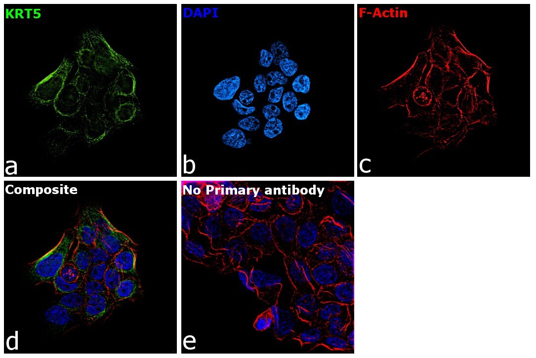
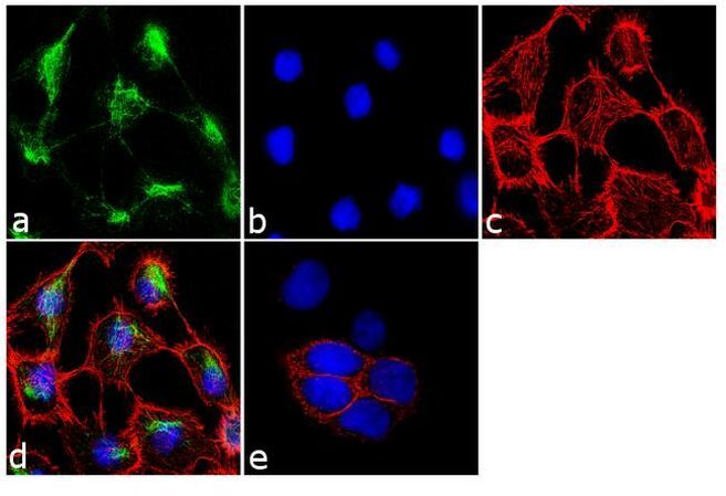
- Immunofluorescence analysis of Cytokeratin 5 was performed using 70% confluent log phase A-431 cells. The cells were fixed with 4% paraformaldehyde for 10 minutes, permeabilized with 0.1% Triton™ X-100 for 10 minutes, and blocked with 1% BSA for 1 hour at room temperature. The cells were labeled with Cytokeratin 5 (XM26) Mouse Monoclonal Antibody (Product # MA5-12596) at 1:250 dilution in 0.1% BSA and incubated for 3 hours at room temperature and then labeled with Goat anti-Mouse IgG (H+L) Superclonal™ Secondary Antibody, Alexa Fluor® 488 conjugate (Product # A28175) at a dilution of 1:2000 for 45 minutes at room temperature (Panel a: green). Nuclei (Panel b: blue) were stained with SlowFade® Gold Antifade Mountant with DAPI (Product # S36938). F-actin (Panel c: red) was stained with Rhodamine Phalloidin (Product # R415, 1:300). Panel d represents the merged image showing cytoplasmic localization. Panel e shows the no primary antibody control. The images were captured at 60X magnification.

- Submitted by
- Invitrogen Antibodies (provider)
- Main image
- Experimental details
- Immunofluorescence analysis of Cytokeratin 5 was performed using 70% confluent log phase A-431 cells. The cells were fixed with ice-cold acetone at 4°C for 5 minutes and blocked with 2% BSA for 45 minutes at room temperature. The cells were labeled with Cytokeratin 5 Monoclonal Antibody (XM26) (Product # MA5-12596) at 1:200 dilution in 0.1% BSA, incubated at 4 degree celsius overnight and then labeled with Goat anti-Mouse IgG (H+L) Superclonal™ Recombinant Secondary Antibody, Alexa Fluor® 488 conjugate (Product # A28175), (1:2000 dilution), for 45 minutes at room temperature (Panel a: Green). Nuclei (Panel b:Blue) were stained with ProLong™ Diamond Antifade Mountant with DAPI (Product # P36962). F-actin (Panel c: Red) was stained with Rhodamine Phalloidin (Product # R415, 1:300 dilution). Panel d represents the merged image showing cytoskeletal localization. Panel e represents control cells with no primary antibody to assess background. The images were captured at 60X magnification.
- Submitted by
- Invitrogen Antibodies (provider)
- Main image

- Experimental details
- Immunofluorescence analysis of Cytokeratin 5 was performed using 70% confluent log phase A-431 cells. The cells were fixed with 4% paraformaldehyde for 10 minutes, permeabilized with 0.1% Triton™ X-100 for 10 minutes, and blocked with 1% BSA for 1 hour at room temperature. The cells were labeled with Cytokeratin 5 (XM26) Mouse Monoclonal Antibody (Product # MA5-12596) at 1:250 dilution in 0.1% BSA and incubated for 3 hours at room temperature and then labeled with Goat anti-Mouse IgG (H+L) Superclonal™ Secondary Antibody, Alexa Fluor® 488 conjugate (Product # A28175) at a dilution of 1:2000 for 45 minutes at room temperature (Panel a: green). Nuclei (Panel b: blue) were stained with SlowFade® Gold Antifade Mountant with DAPI (Product # S36938). F-actin (Panel c: red) was stained with Rhodamine Phalloidin (Product # R415, 1:300). Panel d represents the merged image showing cytoplasmic localization. Panel e shows the no primary antibody control. The images were captured at 60X magnification.
- Submitted by
- Invitrogen Antibodies (provider)
- Main image

- Experimental details
- Immunofluorescence analysis of Cytokeratin 5 was performed using 70% confluent log phase A-431 cells. The cells were fixed with ice-cold acetone at 4°C for 5 minutes and blocked with 2% BSA for 45 minutes at room temperature. The cells were labeled with Cytokeratin 5 Monoclonal Antibody (XM26) (Product # MA5-12596) at 1:200 dilution in 0.1% BSA, incubated at 4 degree celsius overnight and then labeled with Goat anti-Mouse IgG (H+L) Superclonal™ Recombinant Secondary Antibody, Alexa Fluor® 488 conjugate (Product # A28175), (1:2000 dilution), for 45 minutes at room temperature (Panel a: Green). Nuclei (Panel b:Blue) were stained with ProLong™ Diamond Antifade Mountant with DAPI (Product # P36962). F-actin (Panel c: Red) was stained with Rhodamine Phalloidin (Product # R415, 1:300 dilution). Panel d represents the merged image showing cytoskeletal localization. Panel e represents control cells with no primary antibody to assess background. The images were captured at 60X magnification.

Supportive validation
- Submitted by
- Invitrogen Antibodies (provider)
- Main image
- Experimental details
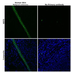
- Immunohistochemical analysis of KRT5 was performed using formalin-fixed paraffin-embedded human skin (malignant melanoma) tissue sections. To expose the target protein, heat-induced epitope retrieval was performed on de-paraffinized sections using eBioscience™ IHC Antigen Retrieval Solution - Low pH (10X) (Product # 00-4955-58) diluted to 1X solution in water in a decloaking chamber at 110 degree Celsius for 15 minutes. Following antigen retrieval, the sections were blocked with 2% normal goat serum in 1X PBS for 45 minutes at room temperature and then probed with or without Cytokeratin 5 Monoclonal Antibody (XM26) (Product # MA5-12596) at 1:100 dilution in 0.1% normal goat serum overnight at 4 degree Celsius in a humidified chamber. Detection was performed using Goat anti-Mouse IgG (H+L) Highly Cross-Adsorbed Secondary Antibody, Alexa Fluor Plus 488 (Product # A32723) at a dilution of 1:2000 in 0.1% normal goat serum for 45 minutes at room temperature. ReadyProbes™ Tissue Autofluorescence Quenching Kit (Product # R37630) was used to quench autofluorescence from the tissues. Nuclei were stained with DAPI (Product # D1306) and the sections were mounted using ProLong™ Glass Antifade Mountant (Product # P36984). The images were captured on EVOS™ M7000 Imaging System (Product # AMF7000) at 20X magnification.

- Submitted by
- Invitrogen Antibodies (provider)
- Main image
- Experimental details
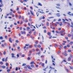
- Formalin-fixed, paraffin-embedded human mesothelioma stained with Keratin 5 antibody using peroxidase-conjugate and AEC chromogen. Note cytoplasmic staining of tumor cells.

- Submitted by
- Invitrogen Antibodies (provider)
- Main image

- Experimental details
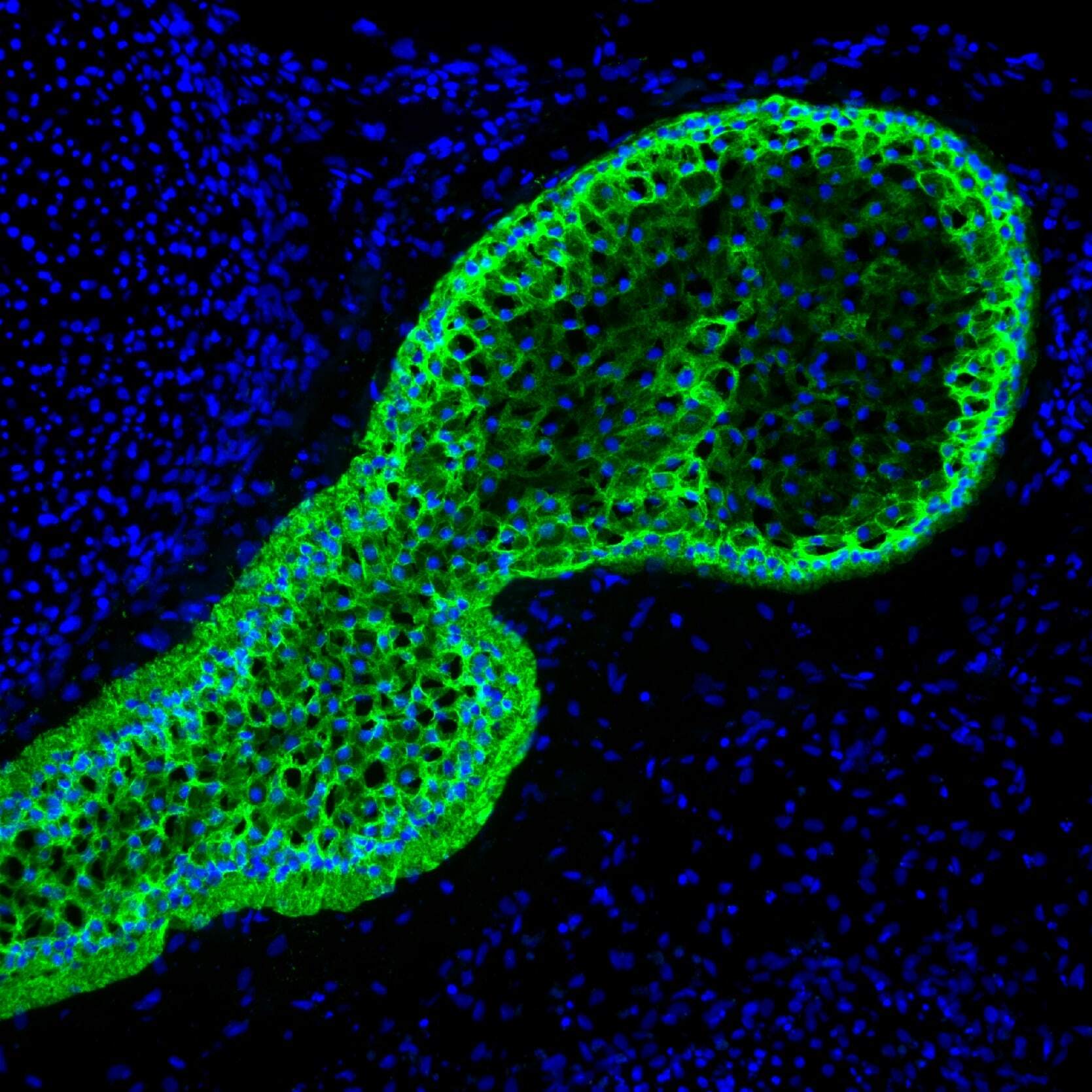
- Immunohistochemistry analysis of Cytokeratin 5 was performed on cryosections of human skin tissue. Tissues were blocked in 10% normal goat serum in 1X PBS containing 0.1% Triton X-100 (1X PBS-T) for 1 hour at room temperature (RT). The tissues were labeled with a Cytokeratin 5 monoclonal antibody (clone XM26, green, Product # MA5-12596) diluted 1:50 in 3% normal goat serum in 1X PBS-T for 1 hour at RT, followed by detection with a Goat anti-Mouse IgG1, Alexa Fluor 488 secondary antibody (Product # A-21121) diluted 1:2000 in 3% normal goat serum in 1X PBS-T for 1 hour at RT. Nuclei (blue) were stained with DAPI, included in ProLong Gold Anti-Fade Mountant (Product # P36931). Images were taken on an inverted microscope at 20X magnification. Data courtesy of Dr. Jiyoon Lee at Indiana University School of Medicine.
Supportive validation
- Submitted by
- Invitrogen Antibodies (provider)
- Main image

- Experimental details
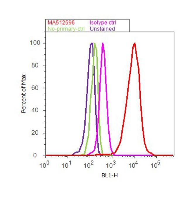
- Flow cytometry analysis of Cytokeratin 5 was done on A-431 cells. Cells were fixed with 70% ethanol for 10 minutes, permeabilized with 0.25% Triton™ X-100 for 20 minutes, and blocked with 5% BSA for 30 minutes at room temperature. Cells were labeled with Cytokeratin 5 Mouse Monoclonal Antibody (MA5-12596, red histogram) or with mouse isotype control (pink histogram) at 3-5 µg/million cells in 2.5% BSA. After incubation at room temperature for 2 hours, the cells were labeled with Alexa Fluor® 488 Rabbit Anti-Mouse Secondary Antibody (A11059) at a dilution of 1:400 for 30 minutes at room temperature. The representative 10,000 cells were acquired and analyzed for each sample using an Attune® Acoustic Focusing Cytometer. The purple histogram represents unstained control cells and the green histogram represents no-primary-antibody control..
Supportive validation
- Submitted by
- Invitrogen Antibodies (provider)
- Main image
- Experimental details
- NULL
- Submitted by
- Invitrogen Antibodies (provider)
- Main image
- Experimental details
- NULL
- Submitted by
- Invitrogen Antibodies (provider)
- Main image
- Experimental details
- NULL

- Submitted by
- Invitrogen Antibodies (provider)
- Main image
- Experimental details
- NULL
- Submitted by
- Invitrogen Antibodies (provider)
- Main image
- Experimental details
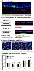
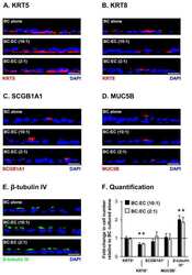
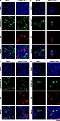
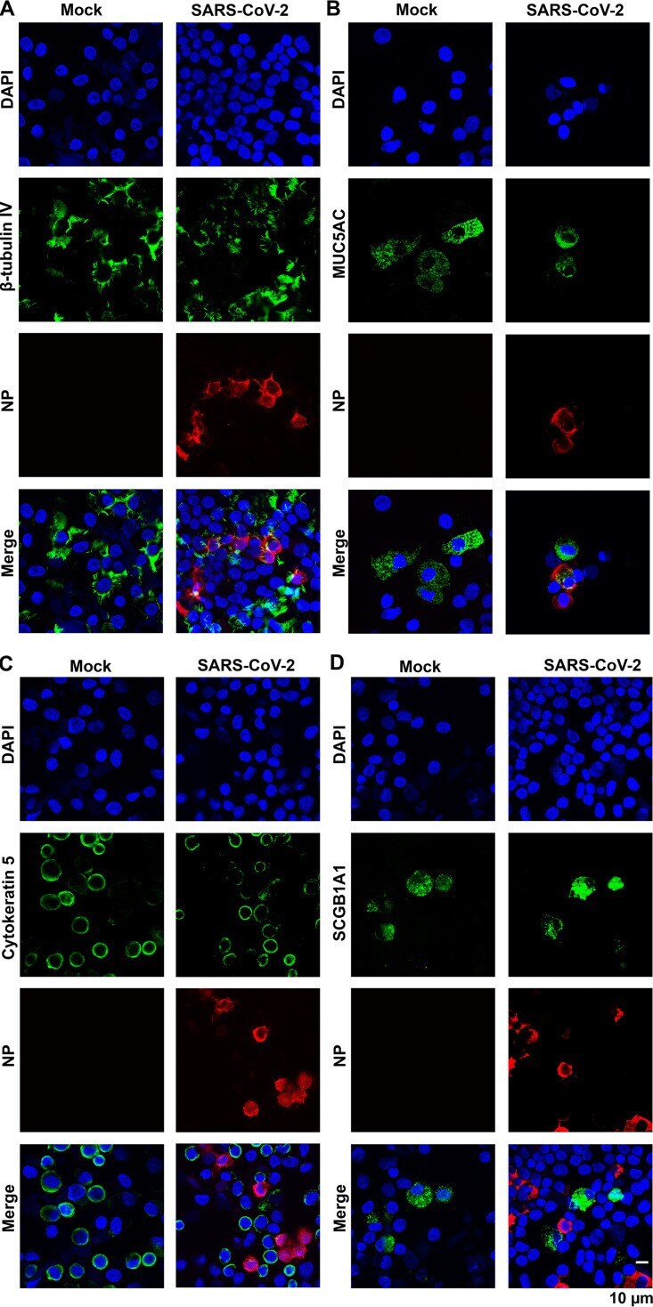
- FIG 7 SARS-CoV-2 infects ciliated and goblet epithelial cells but not basal and club cells. Epithelial cells of the mock- and SARS-CoV-2-infected HAE-ALI B9-20 cultures at 4 dpi (MOI = 0.2) were dissociated from the Transwell insert and cytospun onto slides. The cells on the slides were fixed, permeabilized, and immunostained with anti-NP and together with anti-beta-tubulin IV (A), and anti-MUC5AC (B), anti-cytokeratin 5 (C), and anti-SCGB1A1 (D), respectively. Confocal images were taken at a magnification of x63. Nuclei were stained with DAPI (blue).
- Submitted by
- Invitrogen Antibodies (provider)
- Main image
- Experimental details
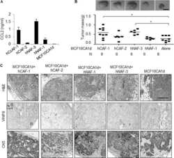
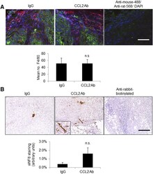
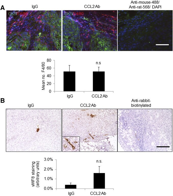
- Figure 5 Delivery of CCL2-neutralizing antibodies did not significantly affect macrophage infiltration or tumor angiogenesis. (A) Primary tumor tissues were co-immunofluorescent stained for CK5 (green) and F4/80 (red). Secondary antibody controls overlaid with 4',6-diamidino-2-phenylindole are shown. (B) Primary tumor tissues were immunostained for vWF8. Magnified insert shows positive staining. Staining was quantified by Image J. Statistical analysis was performed using two-tailed Student's t test. Statistical significance was determined by P < .05. ns = not significant. Mean +- SEM is shown. Scale bar = 200 mum. Figure 5
- Submitted by
- Invitrogen Antibodies (provider)
- Main image
- Experimental details
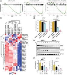
- 3 Figure Treatment of TRb-expressing MDA-MB-468 cells with T 3 represses cytokeratin genes. (A) Gene Set Enrichment Analysis performed between T 3 -treated 468-EV and 468-TRbeta expression analysis revealed Gene Ontology Biological Processes or Hallmark gene sets involving differentially expressed cytokeratins genes (adjusted p value < 1e-7). (B) Heatmap of all expressed KRT genes in 468-EV or 468-TRbeta cells following Vehicle or 10 nM T 3 treatment. (C) Normalized gene expression bar plots of the cytokeratin genes KRT5 and KRT14 from RNA-seq data, n = 3 for each condition. (D) and (E) Protein expression of KRT5 and KRT14 were evaluated by western blot, compared with beta-Actin as a loading control. The blot presented is representative of three independent experiments. **** p < 0.0001. TRbeta, thyroid hormone receptor beta [Color figure can be viewed at wileyonlinelibrary.com ]
- Submitted by
- Invitrogen Antibodies (provider)
- Main image
- Experimental details
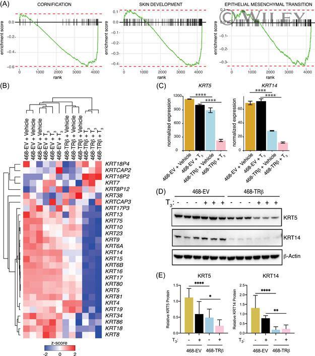
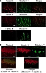
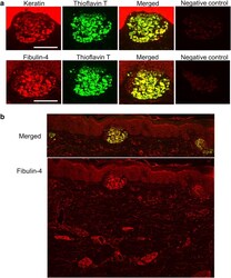
- Keratin 5 and fibulin-4 were found in the amyloid deposits of Bowen's carcinoma and lichen amyloidosis skin. (a) The skin from a Bowen's carcinoma (patient 1) was stained with anti-fibulin-4, anti-keratin 5, and anti-fibrillin-1 antibodies (the left column) together with thioflavin T. Bar = 50 mum. ( b ) Paraffin-embedded sections (patient 2, lichen amyloidosis) were stained with anti-fibulin-4 (red) and anti-keratin 5 (green, data not shown) and with anti-keratin 5 and thioflavin T. Bar = 100 mum.
- Submitted by
- Invitrogen Antibodies (provider)
- Main image
- Experimental details
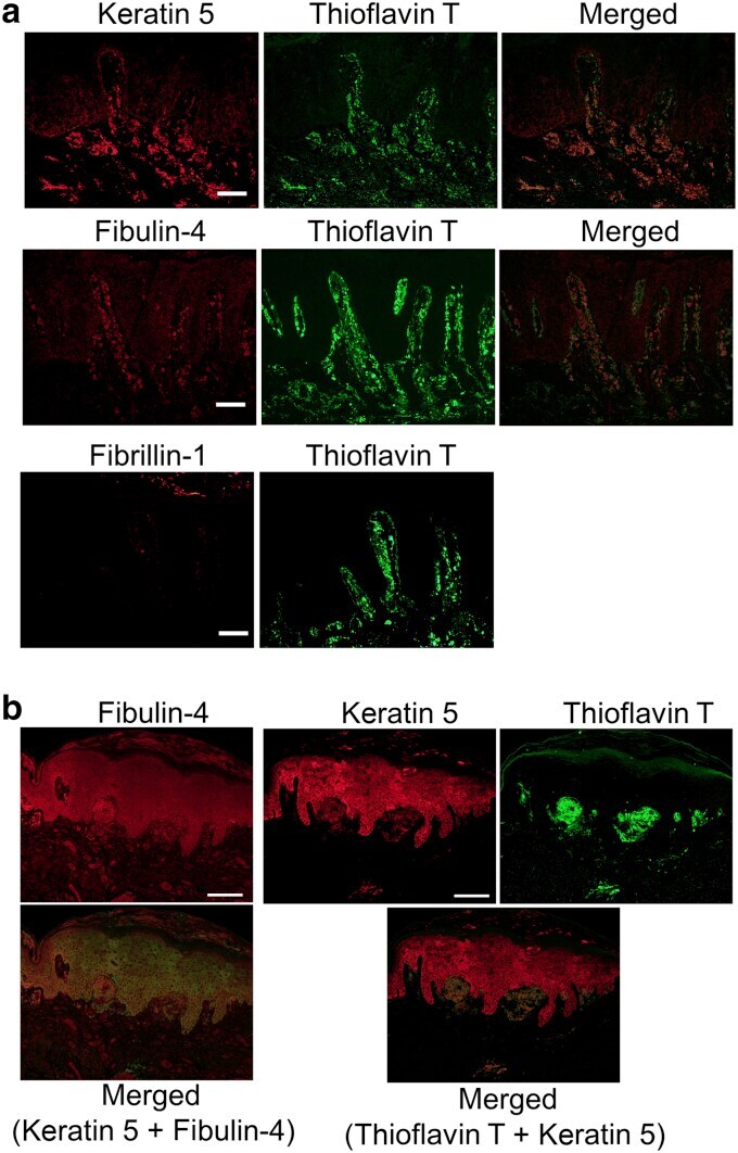
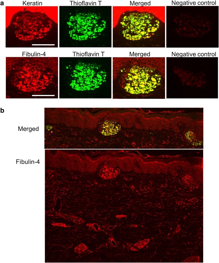
- Fibulin-4 and keratin 5 were found in the amyloid deposits of lichen amyloidosis (patient 3). ( a ) Consecutive frozen sections were stained with anti-keratin 5 or with anti-fibulin-4, and amyloid deposits were detected with thioflavin T. Each merged image is also shown. ( b ) The staining with anti-fibulin-4 in the same section as the second row of a was shown at lower magnification. This image also shows the localization of fibulin-4 in normal human skin. Fibulin-4 localizes in elastic fibers and in the surrounding blood vessels, not only in the papil1ary dermis but also in the reticular dermis. Bar = 100 mum.
- Submitted by
- Invitrogen Antibodies (provider)
- Main image

- Experimental details
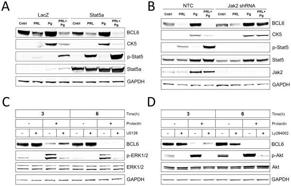
- Figure 5 Prolactin suppression of BCL6 and subsequently CK5 is mediated by the Stat5 pathway (A) Immunoblots of BCL6, CK5, phosphorylated-Stat5 (p-Stat5), Stat5a, and GAPDH protein levels in extracts of T47D cultures exposed to adenovirus carrying either Stat5a or LacZ control for 24 h and subsequently treated with vehicle (Cntrl), Prolactin (PRL), beta-Estradiol (E2), or R5020 (Pg) for 48 h. (B) Immunoblots of BCL6, CK5, phosphorylated-Stat5 (p-Stat5), Stat5, Jak2, and GAPDH protein levels in extracts of T47D cultures treated with non-target shRNA (NTC) or Jak2 shRNA for 48h, followed by addition of vehicle (Cntrl), Prolactin (PRL), beta-Estradiol (E2), or R5020 (Pg) for 48 h. (C) Immunoblots of BCL6, phosphorylated-ERK1/2 (p-ERK1/2), ERK1/2, and GAPDH protein levels in extracts of T47D cultures pre-treated with Pg for 24 h followed by incubation with or without prolactin (PRL) in the presence or absence of Mek inhibitor (U0126) for 3 and 6 h. (D) Immunoblots of BCL6, phosphorylated-Akt (p-Akt), Akt, and GAPDH protein levels in extracts of T47D cultures pre-treated with Pg for 24 h followed by incubation with or without prolactin (PRL) in the presence or absence of PI3K inhibitor (Ly294002) for 3 and 6 h.