Explore
 Validate
 Learn
Learn Western blot
 ELISA
ELISA Immunocytochemistry
Antibody data
- Antibody Data
- Antigen structure
- References [1]
- Comments [0]
- Validations
- Immunocytochemistry [2]
- Immunohistochemistry [2]
- Other assay [2]
Submit
Validation data
Reference
Comment
Report error
- Product number
- MA5-15347 - Provider product page

- Provider
- Invitrogen Antibodies
- Product name
- Cytokeratin 5 Monoclonal Antibody (3E2F1)
- Antibody type
- Monoclonal
- Antigen
- Purifed from natural sources
- Description
- MA5-15347 targets Cytokeratin 5 in indirect ELISA, IHC and WB applications and shows reactivity with Human samples. The MA5-15347 immunogen is purified recombinant fragment of CK5 expressed in E. Coli.
- Reactivity
- Human, Mouse
- Host
- Mouse
- Isotype
- IgG
- Antibody clone number
- 3E2F1
- Vial size
- 100 μL
- Concentration
- Conc. Not Determined
- Storage
- Store at 4°C short term. For long term storage, store at -20°C, avoiding freeze/thaw cycles.
Submitted references Correlation between salivary estrogen levels and oral epithelial cytokeratin 5 expression.
Handajani J, Effendi N, Sosroseno W
F1000Research 2020;9:186
F1000Research 2020;9:186
No comments: Submit comment
Supportive validation
- Submitted by
- Invitrogen Antibodies (provider)
- Main image

- Experimental details
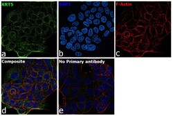
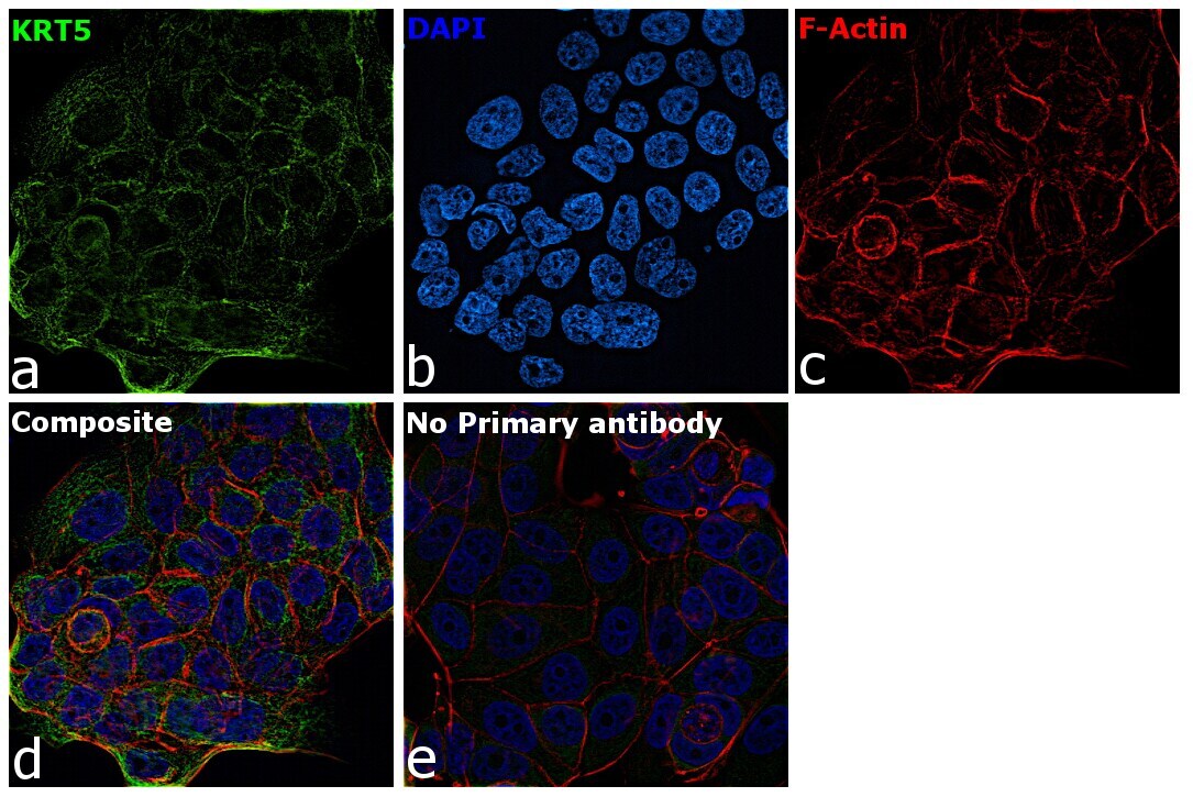
- Immunofluorescence analysis of Cytokeratin 5 was performed using 70% confluent log phase A-431 cells. The cells were fixed with 4% paraformaldehyde for 10 minutes, permeabilized with 0.1% Triton™ X-100 for 10 minutes, and blocked with 2% BSA for 45 minutes at room temperature. The cells were labeled with Cytokeratin 5 Monoclonal Antibody (3E2F1) (Product # MA5-15347) at 1:200 dilution in 0.1% BSA, incubated at 4 degree celsius overnight and then labeled with Goat anti-Mouse IgG (H+L) Superclonal™ Recombinant Secondary Antibody, Alexa Fluor® 488 conjugate (Product # A28175), (1:2000 dilution), for 45 minutes at room temperature (Panel a: Green). Nuclei (Panel b:Blue) were stained with SlowFade® Gold Antifade Mountant with DAPI (Product # S36938). F-actin (Panel c: Red) was stained with Rhodamine Phalloidin (Product # R415, 1:300 dilution). Panel d represents the merged image showing cytoskeletal localization. Panel e represents control cells with no primary antibody to assess background. The images were captured at 60X magnification.

- Submitted by
- Invitrogen Antibodies (provider)
- Main image
- Experimental details
- Immunofluorescence analysis of Cytokeratin 5 was performed using 70% confluent log phase A-431 cells. The cells were fixed with 4% paraformaldehyde for 10 minutes, permeabilized with 0.1% Triton™ X-100 for 10 minutes, and blocked with 2% BSA for 45 minutes at room temperature. The cells were labeled with Cytokeratin 5 Monoclonal Antibody (3E2F1) (Product # MA5-15347) at 1:200 dilution in 0.1% BSA, incubated at 4 degree celsius overnight and then labeled with Goat anti-Mouse IgG (H+L) Superclonal™ Recombinant Secondary Antibody, Alexa Fluor® 488 conjugate (Product # A28175), (1:2000 dilution), for 45 minutes at room temperature (Panel a: Green). Nuclei (Panel b:Blue) were stained with SlowFade® Gold Antifade Mountant with DAPI (Product # S36938). F-actin (Panel c: Red) was stained with Rhodamine Phalloidin (Product # R415, 1:300 dilution). Panel d represents the merged image showing cytoskeletal localization. Panel e represents control cells with no primary antibody to assess background. The images were captured at 60X magnification.
Supportive validation
- Submitted by
- Invitrogen Antibodies (provider)
- Main image
- Experimental details
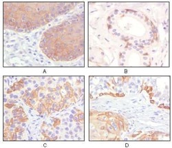
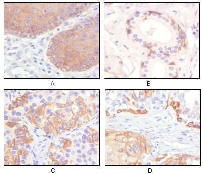
- Immunohistochemical analysis of paraffin-embedded human esophagus epithelium (A), salivary gland basal cell (B), lung squamous cell carcinoma (C), endometrium admosquamous carcinoma (D), showing cytoplasmic and membrane localization using Cytokeratin 5 monoclonal antibody (Product # MA5-15347) followed with DAB staining.
- Submitted by
- Invitrogen Antibodies (provider)
- Main image
- Experimental details
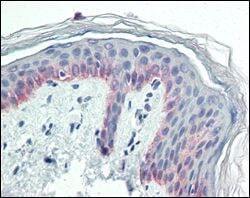
- Immunohistochemical analysis of paraffin-embedded human skin tissues using Cytokeratin 5 monoclonal antibody (Product # MA5-15347).

Supportive validation
- Submitted by
- Invitrogen Antibodies (provider)
- Main image
- Experimental details
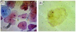
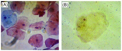
- Figure 1. Palatal epithelial cells stained by Papanicolaou (x400) ( A ) and immunohistologically stained for cytokeratin 5 ( B ) (x400). Basal cells are stained as blue color. Intermediate and superficial are stained as pink and orange color, respectively. Cytokeratin 5 protein is stained as brown color in nucleus and cytoplasm.
- Submitted by
- Invitrogen Antibodies (provider)
- Main image
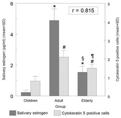
- Experimental details
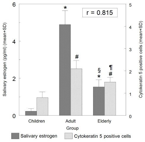
- Figure 2. The correlation between the levels of salivary estrogen and cytokeratin 5-positive oral epithelial cells. *p