 Explore
 Validate
 Learn
Learn Western blot
Antibody data
- Antibody Data
- Antigen structure
- References [14]
- Comments [0]
- Validations
- Western blot [1]
- Immunocytochemistry [1]
- Immunohistochemistry [4]
- Other assay [4]
Submit
Validation data
Reference
Comment
Report error
- Product number
- MA5-14473 - Provider product page

- Provider
- Invitrogen Antibodies
- Product name
- Cytokeratin 5 Monoclonal Antibody (EP1601Y)
- Antibody type
- Monoclonal
- Antigen
- Synthetic peptide
- Description
- MA5-14473 targets Cytokeratin 5 in IHC (P) applications and shows reactivity with Human samples.
- Antibody clone number
- EP1601Y
- Concentration
- Conc. Not Determined
Submitted references Malfunction of airway basal stem cells plays a crucial role in pathophysiology of tracheobronchopathia osteoplastica.
PI3Kδ Sustains Keratinocyte Hyperproliferation and Epithelial Inflammation: Implications for a Topically Druggable Target in Psoriasis.
Temporospatial Expression of Fgfr1 and 2 During Lung Development, Homeostasis, and Regeneration.
Alveolar Differentiation Potency of Human Distal Airway Stem Cells Is Associated with Pulmonary Pathological Conditions.
A Preclinical Model for Studying Herpes Simplex Virus Infection.
Hippo signaling promotes lung epithelial lineage commitment by curbing Fgf10 and β-catenin signaling.
Fgf10-Hippo Epithelial-Mesenchymal Crosstalk Maintains and Recruits Lung Basal Stem Cells.
Molecular subtypes of urothelial carcinoma are defined by specific gene regulatory systems.
p63(+)Krt5(+) distal airway stem cells are essential for lung regeneration.
A novel blueprint for 'top down' differentiation defines the cervical squamocolumnar junction during development, reproductive life, and neoplasia.
A discrete population of squamocolumnar junction cells implicated in the pathogenesis of cervical cancer.
A molecular taxonomy for urothelial carcinoma.
An immunohistochemical study of the origin of the solid strand in syringoma, using carcinoembryonic antigen, epithelial membrane antigen, and cytokeratin 5.
Characterization of the expression of cytokeratins 5, 8, and 14 in mouse thymic epithelial cells during thymus regeneration following acute thymic involution.
Hong Y, Shan S, Gu Y, Huang H, Zhang Q, Han Y, Dong Y, Liu Z, Huang M, Ren T
Nature communications 2022 Mar 14;13(1):1309
Nature communications 2022 Mar 14;13(1):1309
PI3Kδ Sustains Keratinocyte Hyperproliferation and Epithelial Inflammation: Implications for a Topically Druggable Target in Psoriasis.
Mercurio L, Morelli M, Scarponi C, Scaglione GL, Pallotta S, Albanesi C, Madonna S
Cells 2021 Oct 2;10(10)
Cells 2021 Oct 2;10(10)
Temporospatial Expression of Fgfr1 and 2 During Lung Development, Homeostasis, and Regeneration.
Yuan T, Klinkhammer K, Lyu H, Gao S, Yuan J, Hopkins S, Zhang JS, De Langhe SP
Frontiers in pharmacology 2020;11:120
Frontiers in pharmacology 2020;11:120
Alveolar Differentiation Potency of Human Distal Airway Stem Cells Is Associated with Pulmonary Pathological Conditions.
Wang Y, Lu Y, Wu Y, Sun Y, Zhou Y, Ma Q, Zheng Y, Yu Q, Cao Y, Chen G, Zhang T, Dai X, Ren T, Ma Y, Zuo W
Stem cells international 2019;2019:7123078
Stem cells international 2019;2019:7123078
A Preclinical Model for Studying Herpes Simplex Virus Infection.
Tajpara P, Mildner M, Schmidt R, Vierhapper M, Matiasek J, Popow-Kraupp T, Schuster C, Elbe-Bürger A
The Journal of investigative dermatology 2019 Mar;139(3):673-682
The Journal of investigative dermatology 2019 Mar;139(3):673-682
Hippo signaling promotes lung epithelial lineage commitment by curbing Fgf10 and β-catenin signaling.
Volckaert T, Yuan T, Yuan J, Boateng E, Hopkins S, Zhang JS, Thannickal VJ, Fässler R, De Langhe SP
Development (Cambridge, England) 2019 Jan 16;146(2)
Development (Cambridge, England) 2019 Jan 16;146(2)
Fgf10-Hippo Epithelial-Mesenchymal Crosstalk Maintains and Recruits Lung Basal Stem Cells.
Volckaert T, Yuan T, Chao CM, Bell H, Sitaula A, Szimmtenings L, El Agha E, Chanda D, Majka S, Bellusci S, Thannickal VJ, Fässler R, De Langhe SP
Developmental cell 2017 Oct 9;43(1):48-59.e5
Developmental cell 2017 Oct 9;43(1):48-59.e5
Molecular subtypes of urothelial carcinoma are defined by specific gene regulatory systems.
Eriksson P, Aine M, Veerla S, Liedberg F, Sjödahl G, Höglund M
BMC medical genomics 2015 May 26;8:25
BMC medical genomics 2015 May 26;8:25
p63(+)Krt5(+) distal airway stem cells are essential for lung regeneration.
Zuo W, Zhang T, Wu DZ, Guan SP, Liew AA, Yamamoto Y, Wang X, Lim SJ, Vincent M, Lessard M, Crum CP, Xian W, McKeon F
Nature 2015 Jan 29;517(7536):616-20
Nature 2015 Jan 29;517(7536):616-20
A novel blueprint for 'top down' differentiation defines the cervical squamocolumnar junction during development, reproductive life, and neoplasia.
Herfs M, Vargas SO, Yamamoto Y, Howitt BE, Nucci MR, Hornick JL, McKeon FD, Xian W, Crum CP
The Journal of pathology 2013 Feb;229(3):460-8
The Journal of pathology 2013 Feb;229(3):460-8
A discrete population of squamocolumnar junction cells implicated in the pathogenesis of cervical cancer.
Herfs M, Yamamoto Y, Laury A, Wang X, Nucci MR, McLaughlin-Drubin ME, Münger K, Feldman S, McKeon FD, Xian W, Crum CP
Proceedings of the National Academy of Sciences of the United States of America 2012 Jun 26;109(26):10516-21
Proceedings of the National Academy of Sciences of the United States of America 2012 Jun 26;109(26):10516-21
A molecular taxonomy for urothelial carcinoma.
Sjödahl G, Lauss M, Lövgren K, Chebil G, Gudjonsson S, Veerla S, Patschan O, Aine M, Fernö M, Ringnér M, Månsson W, Liedberg F, Lindgren D, Höglund M
Clinical cancer research : an official journal of the American Association for Cancer Research 2012 Jun 15;18(12):3377-86
Clinical cancer research : an official journal of the American Association for Cancer Research 2012 Jun 15;18(12):3377-86
An immunohistochemical study of the origin of the solid strand in syringoma, using carcinoembryonic antigen, epithelial membrane antigen, and cytokeratin 5.
Kim BC, Park EJ, Kwon IH, Cho HJ, Park HR, Kim KH, Kim KJ
International journal of dermatology 2012 Jul;51(7):817-22
International journal of dermatology 2012 Jul;51(7):817-22
Characterization of the expression of cytokeratins 5, 8, and 14 in mouse thymic epithelial cells during thymus regeneration following acute thymic involution.
Lee EN, Park JK, Lee JR, Oh SO, Baek SY, Kim BS, Yoon S
Anatomy & cell biology 2011 Mar;44(1):14-24
Anatomy & cell biology 2011 Mar;44(1):14-24
No comments: Submit comment
Supportive validation
- Submitted by
- Invitrogen Antibodies (provider)
- Main image
- Experimental details
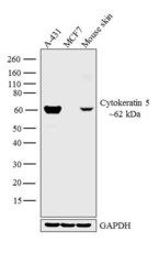
- Western blot analysis was performed on whole cell extracts (30 µg lysate) of A-431 (Lane 1), MCF7 (Lane 2) and tissue extracts (30 µg lysate) of Mouse skin. The blot was probed with Anti-Cytokeratin 5 Monoclonal Antibody (Product # MA5-14473, 1:500 dilution) and detected by chemiluminescence using Goat anti-Rabbit IgG (H+L) Superclonal™ Secondary Antibody, HRP conjugate (Product # A27036, 0.25 µg/mL, 1:4000 dilution). A 62 kDa band corresponding to Cytokeratin 5 was observed across the cell lines tested. Known quantity of protein samples were electrophoresed using Novex® NuPAGE® 4-12 % Bis-Tris gel (Product # NP0321BOX), XCell SureLock™ Electrophoresis System (Product # EI0002) and Novex® Sharp Pre-Stained Protein Standard (Product # LC5800). Resolved proteins were then transferred onto a nitrocellulose membrane with iBlot® 2 Dry Blotting System (Product # IB21001). The membrane was probed with the relevant primary and secondary Antibody following blocking with 5 % skimmed milk. Chemiluminescent detection was performed using Pierce™ ECL Western Blotting Substrate (Product # 32106).

Supportive validation
- Submitted by
- Invitrogen Antibodies (provider)
- Main image

- Experimental details
- Immunofluorescence analysis of Cytokeratin 5 was performed using 70% confluent log phase A-431 cells. The cells were fixed with 4% paraformaldehyde for 10 minutes, permeabilized with 0.1% Triton™ X-100 for 10 minutes, and blocked with 1% BSA for 1 hour at room temperature. The cells were labeled with Cytokeratin 5 Monoclonal Antibody (Product # MA5-14473) at 5µg/mL in 0.1% BSA and incubated for 3 hours at room temperature and then labeled with Goat anti-Rabbit IgG (H+L) Superclonal™ Secondary Antibody, Alexa Fluor® 488 conjugate (Product # A27034) at a dilution of 1:2000 for 45 minutes at room temperature (Panel a: green). Nuclei (Panel b: blue) were stained with SlowFade® Gold Antifade Mountant with DAPI (Product # S36938). F-actin (Panel c: red) was stained with Rhodamine Phalloidin (Product # R415, 1:300). Panel d represents the merged image showing cytoplasmic localization. Panel e shows the no primary antibody control. The images were captured at 60X magnification.

Supportive validation
- Submitted by
- Invitrogen Antibodies (provider)
- Main image

- Experimental details
- Formalin-fixed, paraffin-embedded human mesothelioma stained with Keratin 5 using peroxidase-conjugate and DAB chromogen. Note cytoplasmic staining.

- Submitted by
- Invitrogen Antibodies (provider)
- Main image
- Experimental details
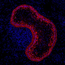
- Immunohistochemistry analysis of Cytokeratin 5 was performed on cryosections of human skin tissue. Tissues were blocked in 10% normal goat serum in 1X PBS containing 0.1% Triton X-100 (1X PBS-T) for 1 hour at room temperature (RT). The tissues were labeled with a Cytokeratin 5 monoclonal antibody (clone EP1601Y, red, Product # MA5-14473) diluted 1:50 in 3% normal goat serum in 1X PBS-T for 1 hour at RT, followed by detection with a Goat anti-Rabbit IgG (H+L), Alexa Fluor 568 secondary antibody (Product # A-11036) diluted 1:2000 in 3% normal goat serum in 1X PBS-T for 1 hour at RT. Nuclei (blue) were stained with DAPI, included in ProLong Gold Anti-Fade Mountant (Product # P36931). Images were taken on an inverted microscope at 20X magnification. Data courtesy of Dr. Jiyoon Lee at Indiana University School of Medicine.

- Submitted by
- Invitrogen Antibodies (provider)
- Main image

- Experimental details
- Immunohistochemical analysis of KRT5 was performed using formalin-fixed paraffin-embedded human skin (malignant melanoma) tissue sections. To expose the target protein, heat-induced epitope retrieval was performed on de-paraffinized sections using eBioscience™ IHC Antigen Retrieval Solution - Low pH (10X) (Product # 00-4955-58) diluted to 1X solution in water in a decloaking chamber at 110 degree Celsius for 15 minutes. Following antigen retrieval, the sections were blocked with 2% normal goat serum in 1X PBS for 45 minutes at room temperature and then probed with or without Cytokeratin 5 Monoclonal Antibody (EP1601Y) (Product # MA5-14473) at 1:100 dilution in 0.1% normal goat serum overnight at 4 degree Celsius in a humidified chamber. Detection was performed using Goat anti-Rabbit IgG (H+L) Highly Cross-Adsorbed Secondary Antibody, Alexa Fluor Plus 488 (Product # A32731) at a dilution of 1:2000 in 0.1% normal goat serum for 45 minutes at room temperature. ReadyProbes™ Tissue Autofluorescence Quenching Kit (Product # R37630) was used to quench autofluorescence from the tissues. Nuclei were stained with DAPI (Product # D1306) and the sections were mounted using ProLong™ Glass Antifade Mountant (Product # P36984). The images were captured on EVOS™ M7000 Imaging System (Product # AMF7000) at 20X magnification.

- Submitted by
- Invitrogen Antibodies (provider)
- Main image

- Experimental details
- Immunohistochemical analysis of KRT5 was performed using formalin-fixed paraffin-embedded mouse skin and mouse brain tissue sections. To expose the target protein, heat-induced epitope retrieval was performed on de-paraffinized sections using eBioscience™ IHC Antigen Retrieval Solution - Low pH (10X) (Product # 00-4955-58) diluted to 1X solution in water in a decloaking chamber at 90 degree Celsius for 20 minutes. Following antigen retrieval, the sections were blocked with 2% normal goat serum in 1X PBS for 45 minutes at room temperature and then probed with Cytokeratin 5 Monoclonal Antibody (EP1601Y) (Product # MA5-14473) at 1:100 dilution in 0.1% normal goat serum overnight at 4 degree Celsius in a humidified chamber. Detection was performed using Goat anti-Rabbit IgG (H+L) Highly Cross-Adsorbed Secondary Antibody, Alexa Fluor Plus 488 (Product # A32731) at a dilution of 1:2000 in 0.1% normal goat serum for 45 minutes at room temperature. Nuclei were stained with DAPI (Product # D1306) and the sections were mounted using ProLong™ Glass Antifade Mountant (Product # P36984). The images were captured on EVOS™ M7000 Imaging System (Product # AMF7000) at 20X magnification and externally deconvoluted (D.Sage et al. / Methods 115 (2017) 28–41).

Supportive validation
- Submitted by
- Invitrogen Antibodies (provider)
- Main image

- Experimental details
- NULL

- Submitted by
- Invitrogen Antibodies (provider)
- Main image

- Experimental details
- NULL

- Submitted by
- Invitrogen Antibodies (provider)
- Main image

- Experimental details
- NULL

- Submitted by
- Invitrogen Antibodies (provider)
- Main image

- Experimental details
- Figure 1 PI3Kdelta expression is up-regulated in skin of psoriatic patients and in proliferating psoriatic keratinocytes activated by pro-inflammatory cytokines. ( A ) In GSE13355 dataset, the raw data from 180 microarrays were processed using the robust multichip average (RMA) method. The resulting expression values of the PI3Kdelta, alpha, and beta isoforms enzymes in healthy control (Healthy, n = 64), non-lesional (NLS, n = 58) and lesional (LS, n = 58) psoriatic skin tissues were obtained from RNA-seq dataset (GSE13355). Datasets were obtained from the transcriptome analysis of whole biopsies from lesional (LS) and non-lesional (NLS) psoriatic skin. Data are expressed as mean +- SD. Statistical significance was assessed by paired Student's t test, * p
