Explore
 Validate
 Learn
Learn Western blot
 Immunocytochemistry
Antibody data
- Antibody Data
- Antigen structure
- References [1]
- Comments [0]
- Validations
- Immunocytochemistry [2]
- Immunohistochemistry [5]
- Other assay [1]
Submit
Validation data
Reference
Comment
Report error
- Product number
- PA5-53954 - Provider product page

- Provider
- Invitrogen Antibodies
- Product name
- Aquaporin 1 Polyclonal Antibody
- Antibody type
- Polyclonal
- Antigen
- Recombinant protein fragment
- Description
- Immunogen sequence: PRSSDLTDRV KVWTSGQVEE YDLDADDINS RVEMKPK Highest antigen sequence identity to the following orthologs: Mouse - 95%, Rat - 95%.
- Reactivity
- Human
- Host
- Rabbit
- Isotype
- IgG
- Vial size
- 100 μL
- Concentration
- 0.10 mg/mL
- Storage
- Store at 4°C short term. For long term storage, store at -20°C, avoiding freeze/thaw cycles.
Submitted references Hamster organotypic kidney culture model of early-stage SARS-CoV-2 infection highlights a two-step renal susceptibility.
Shyfrin SR, Ferren M, Perrin-Cocon L, Espi M, Charmetant X, Brailly M, Decimo D, Iampietro M, Canus L, Horvat B, Lotteau V, Vidalain PO, Thaunat O, Mathieu C
Journal of tissue engineering 2022 Jan-Dec;13:20417314221122130
Journal of tissue engineering 2022 Jan-Dec;13:20417314221122130
No comments: Submit comment
Supportive validation
- Submitted by
- Invitrogen Antibodies (provider)
- Main image
- Experimental details
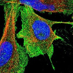
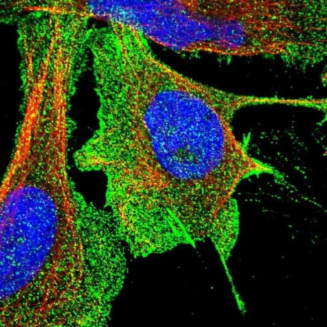
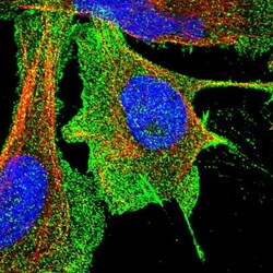
- Immunofluorescent staining of Aquaporin 1 in human cell line U-2 OS shows positivity in plasma membrane. Samples were probed using an Aquaporin 1 Polyclonal Antibody (Product # PA5-53954).
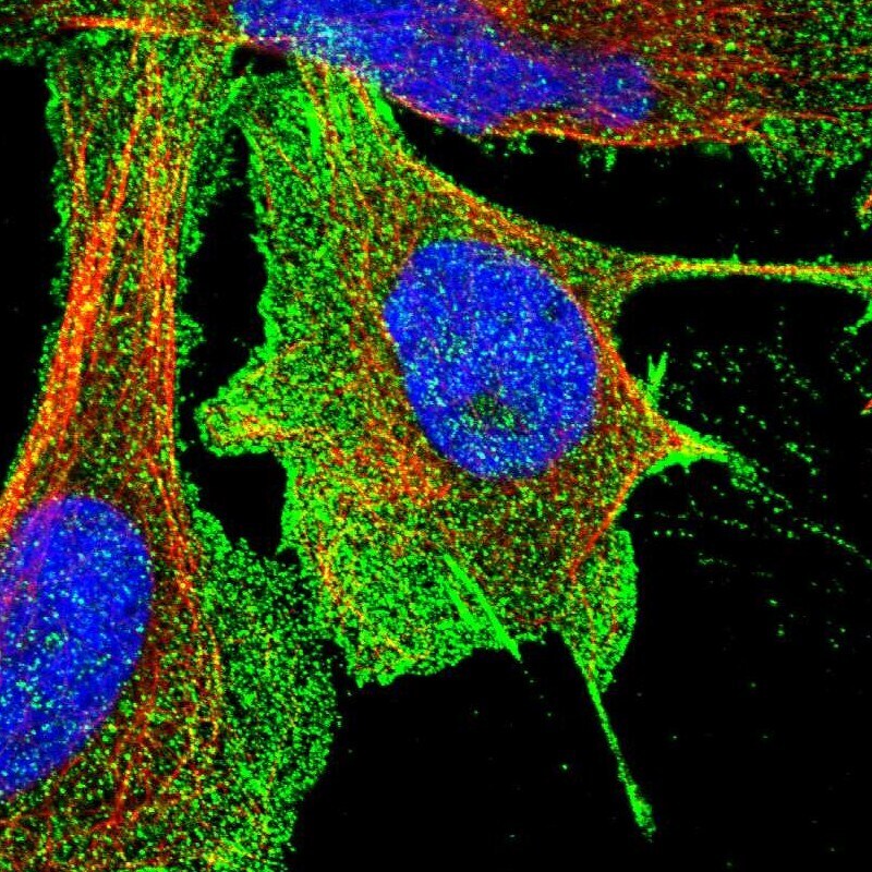
- Submitted by
- Invitrogen Antibodies (provider)
- Main image
- Experimental details
- Immunofluorecent analysis of Aquaporin 1 in human cell line U-2 OS using Aquaporin 1 Polyclonal Antibody (Product # PA5-53954). Staining shows localization to plasma membrane.
Supportive validation
- Submitted by
- Invitrogen Antibodies (provider)
- Main image

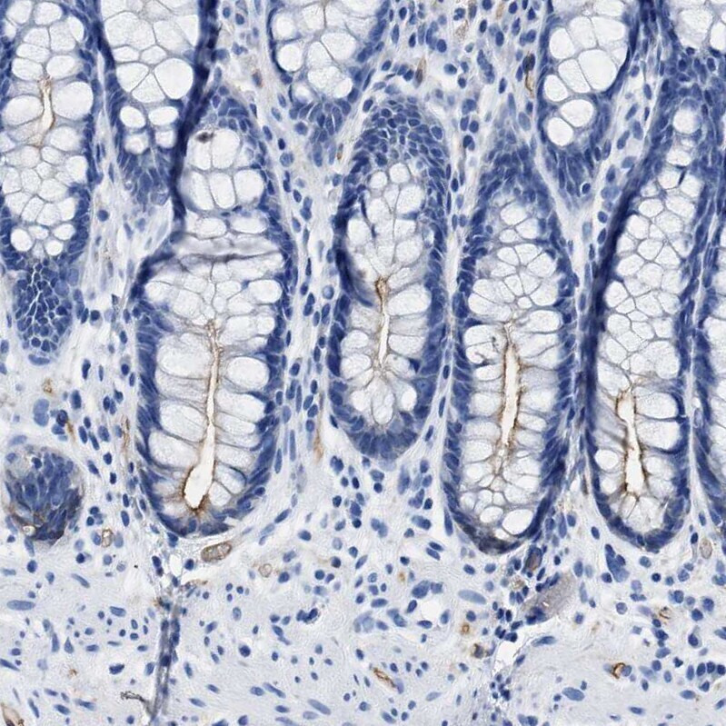
- Experimental details
- Immunohistochemical staining of Aquaporin 1 in human rectum using Aquaporin 1 Polyclonal Antibody (Product # PA5-53954) shows moderate positivity in apical membranes in glandular cells.
- Submitted by
- Invitrogen Antibodies (provider)
- Main image
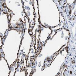
- Experimental details
- Immunohistochemical staining of Aquaporin 1 in human lung using Aquaporin 1 Polyclonal Antibody (Product # PA5-53954) shows strong membranous positivity in endothelial cells.
- Submitted by
- Invitrogen Antibodies (provider)
- Main image
- Experimental details
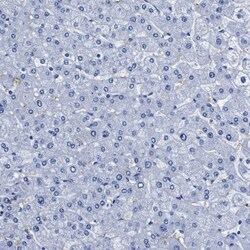
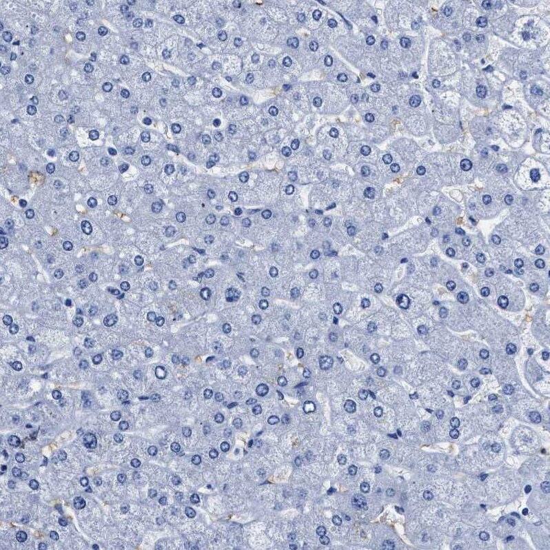
- Immunohistochemical staining of Aquaporin 1 in human liver using Aquaporin 1 Polyclonal Antibody (Product # PA5-53954) shows no positivity in hepatocytes as expected.
- Submitted by
- Invitrogen Antibodies (provider)
- Main image
- Experimental details
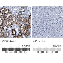
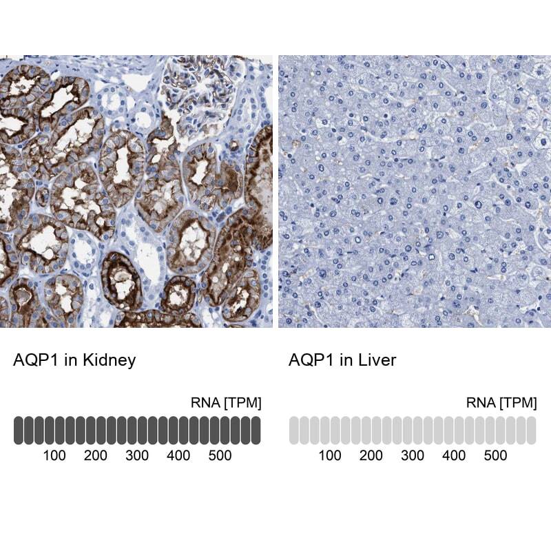
- Immunohistochemical staining of Aquaporin 1 in human kidney and liver tissues using Aquaporin 1 Polyclonal Antibody (Product # PA5-53954). Corresponding AQP1 RNA-seq data are presented for the same tissues.
- Submitted by
- Invitrogen Antibodies (provider)
- Main image
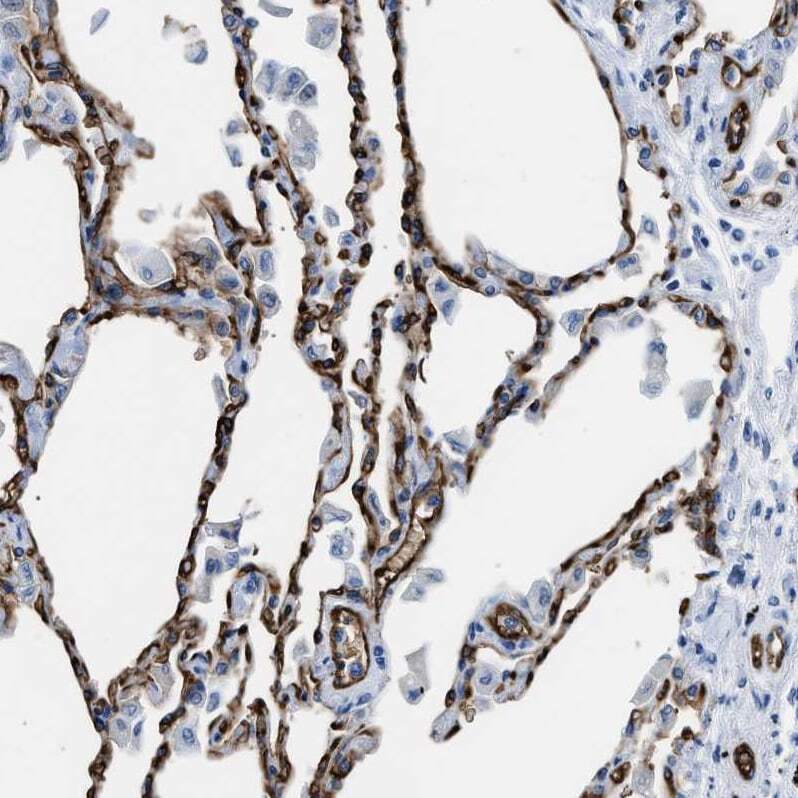
- Experimental details
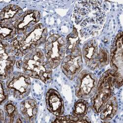
- Immunohistochemical staining of Aquaporin 1 in human kidney using Aquaporin 1 Polyclonal Antibody (Product # PA5-53954) shows moderate to strong positivity in apical membranes in cells in tubules.

Supportive validation
- Submitted by
- Invitrogen Antibodies (provider)
- Main image
- Experimental details
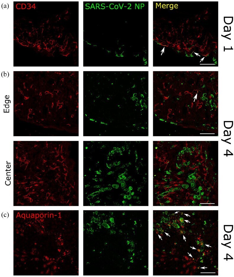
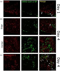
- Figure 4. Tropism and dissemination of SARS-CoV-2 in hamster organotypic kidney cultures. Organotypic kidney cultures (OKC) were infected with 1000 pfu of wild-type SARS-CoV-2 and fixed in 4% paraformaldehyde at 1 or 4 days post infection (dpi). (a and b) OKC sections stained against SARS-CoV-2 nucleoprotein (NP) and CD34 (marker of endothelial cells) at day 1 and 4 post infection ((a and b), respectively). (a) is showing the edge of the slice. (b) is showing both the edge and the center of the slice, demonstrating the spread of infection toward the center. (c) OKC sections stained against SARS-CoV-2 NP and (c) aquaporin-1 (marker of proximal tubular epithelial cells). Immunofluorescence images were acquired using confocal microscopy and is representative of three independent experiments. Colocalization of cell type markers (red) with SARS-CoV-2 NP (green) is denoted with arrows. Scalebar = 100 um.