Explore
 Validate
 Learn
Learn Western blot
 Immunocytochemistry
Antibody data
- Antibody Data
- Antigen structure
- References [7]
- Comments [0]
- Validations
- Western blot [1]
Submit
Validation data
Reference
Comment
Report error
- Product number
- PA1010 - Provider product page

- Provider
- Boster Biological Technology
- Product name
- Anti-Aquaporin 1/AQP1 Antibody
- Antibody type
- Polyclonal
- Description
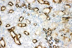
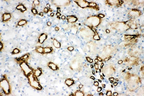
- Polyclonal antibody for AQP1 detection. Host: Rabbit.Size: 100μg/vial. Tested applications: IHC-P. Reactive species: Human. AQP1 information: Molecular Weight: 28526 MW; Subcellular Localization: Cell membrane ; Multi-pass membrane protein ; Tissue Specificity: Detected in erythrocytes (at protein level). Expressed in a number of tissues including erythrocytes, renal tubules, retinal pigment epithelium, heart, lung, skeletal muscle, kidney and pancreas. Weakly expressed in brain, placenta and liver.
- Reactivity
- Human, Mouse, Rat
- Host
- Rabbit
- Vial size
- 100μg/vial
- Concentration
- Add 0.2ml of distilled water will yield a concentration of 500ug/ml.
- Storage
- At -20°C for one year. After reconstitution, at 4°C for one month. It can also be aliquoted and stored frozen at -20°C for a longer time. Avoid repeated freezing and thawing.
- Handling
- Add 0.2ml of distilled water will yield a concentration of 500ug/ml.
Submitted references Tanshinone IIA increased amniotic fluid volume through down-regulating placental AQPs expression via inhibiting the activity of GSK-3β.
GSK-3β inhibitor TDZD-8 prevents reduction of aquaporin-1 expression via activating autophagy under renal ischemia reperfusion injury.
MEF2C/miR-133a-3p.1 circuit-stabilized AQP1 expression maintains endothelial water homeostasis.
Combination exposure of melamine and cyanuric acid is associated with polyuria and activation of NLRP3 inflammasome in rats.
Expression pattern of aquaporins in patients with primary nephrotic syndrome with edema.
Reconstruction of a tissue-engineered cornea with porcine corneal acellular matrix as the scaffold.
Aquaporin-1 expression and angiogenesis in rabbit chronic myocardial ischemia is decreased by acetazolamide.
Shao H, Pan S, Lan Y, Chen X, Dai D, Peng L, Hua Y
Cell and tissue research 2022 Sep;389(3):547-558
Cell and tissue research 2022 Sep;389(3):547-558
GSK-3β inhibitor TDZD-8 prevents reduction of aquaporin-1 expression via activating autophagy under renal ischemia reperfusion injury.
Liu Q, Kong Y, Guo X, Liang B, Xie H, Hu S, Han M, Zhao X, Feng P, Lyu Q, Dong W, Liang X, Wang W, Li C
FASEB journal : official publication of the Federation of American Societies for Experimental Biology 2021 Aug;35(8):e21809
FASEB journal : official publication of the Federation of American Societies for Experimental Biology 2021 Aug;35(8):e21809
MEF2C/miR-133a-3p.1 circuit-stabilized AQP1 expression maintains endothelial water homeostasis.
Jiang Y, Ma R, Zhao Y, Li GJ, Wang AK, Lin WL, Lan XM, Zhong SY, Cai JH
FEBS letters 2019 Sep;593(18):2566-2573
FEBS letters 2019 Sep;593(18):2566-2573
Combination exposure of melamine and cyanuric acid is associated with polyuria and activation of NLRP3 inflammasome in rats.
Wang F, Liu Q, Jin L, Hu S, Luo R, Han M, Zhai Y, Wang W, Li C
American journal of physiology. Renal physiology 2018 Aug 1;315(2):F199-F210
American journal of physiology. Renal physiology 2018 Aug 1;315(2):F199-F210
Expression pattern of aquaporins in patients with primary nephrotic syndrome with edema.
Wang Y, Bu J, Zhang Q, Chen K, Zhang J, Bao X
Molecular medicine reports 2015 Oct;12(4):5625-32
Molecular medicine reports 2015 Oct;12(4):5625-32
Reconstruction of a tissue-engineered cornea with porcine corneal acellular matrix as the scaffold.
Fu Y, Fan X, Chen P, Shao C, Lu W
Cells, tissues, organs 2010;191(3):193-202
Cells, tissues, organs 2010;191(3):193-202
Aquaporin-1 expression and angiogenesis in rabbit chronic myocardial ischemia is decreased by acetazolamide.
Ran X, Wang H, Chen Y, Zeng Z, Zhou Q, Zheng R, Sun J, Wang B, Lv X, Liang Y, Zhang K, Liu W
Heart and vessels 2010 May;25(3):237-47
Heart and vessels 2010 May;25(3):237-47
No comments: Submit comment
Supportive validation
- Submitted by
- Boster Biological Technology (provider)
- Main image
- Experimental details
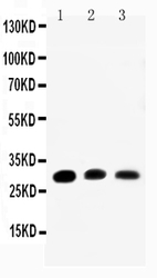
- Western blot analysis of Aquaporin 1 using anti- Aquaporin 1 antibody (PA1010). Electrophoresis was performed on a 5-20% SDS-PAGE gel at 70V (Stacking gel) / 90V (Resolving gel) for 2-3 hours. The sample well of each lane was loaded with 50ug of sample under reducing conditions. Lane 1: Rat Kidney Tissue Lysate, Lane 2: Rat Lung Tissue Lysate, Lane 3: SMMC Cell Lysate. After Electrophoresis, proteins were transferred to a Nitrocellulose membrane at 150mA for 50-90 minutes. Blocked the membrane with 5% Non-fat Milk/ TBS for 1.5 hour at RT. The membrane was incubated with rabbit anti- Aquaporin 1 antigen affinity purified polyclonal antibody (Catalog # PA1010) at 0.5 μg/mL overnight at 4°C, then washed with TBS-0.1%Tween 3 times with 5 minutes each and probed with a goat anti-rabbit IgG-HRP secondary antibody at a dilution of 1:10000 for 1.5 hour at RT. The signal is developed using an Enhanced Chemiluminescent detection (ECL) kit (Catalog # EK1002) with Tanon 5200 system. A specific band was detected for Aquaporin 1 at approximately 28KD. The expected band size for Aquaporin 1 is at 28KD.
- Additional image