Explore
 Validate
 Learn
Learn Western blot
 Immunocytochemistry
Antibody data
- Antibody Data
- Antigen structure
- References [3]
- Comments [0]
- Validations
- Western blot [1]
- Immunocytochemistry [1]
- Immunohistochemistry [1]
Submit
Validation data
Reference
Comment
Report error
- Product number
- HPA019206 - Provider product page

- Provider
- Atlas Antibodies
- Proper citation
- Atlas Antibodies Cat#HPA019206, RRID:AB_1844965
- Product name
- Anti-AQP1
- Antibody type
- Polyclonal
- Description
- Polyclonal Antibody against Human AQP1, Gene description: aquaporin 1 (Colton blood group), Alternative Gene Names: CHIP28, CO, Validated applications: ICC, IHC, WB, Uniprot ID: P29972, Storage: Store at +4°C for short term storage. Long time storage is recommended at -20°C.
- Reactivity
- Human
- Host
- Rabbit
- Conjugate
- Unconjugated
- Isotype
- IgG
- Vial size
- 100 µl
- Concentration
- 0.1 mg/ml
- Storage
- Store at +4°C for short term storage. Long time storage is recommended at -20°C.
- Handling
- The antibody solution should be gently mixed before use.
Submitted references Monkeys mutant for PKD1 recapitulate human autosomal dominant polycystic kidney disease
Aquaporin 1 expression is associated with response to adjuvant chemotherapy in stage II and III colorectal cancer.
Autoantibodies Targeting a Collecting Duct–Specific Water Channel in Tubulointerstitial Nephritis
Tsukiyama T, Kobayashi K, Nakaya M, Iwatani C, Seita Y, Tsuchiya H, Matsushita J, Kitajima K, Kawamoto I, Nakagawa T, Fukuda K, Iwakiri T, Izumi H, Itagaki I, Kume S, Maegawa H, Nishinakamura R, Nishio S, Nakamura S, Kawauchi A, Ema M
Nature Communications 2019;10(1)
Nature Communications 2019;10(1)
Aquaporin 1 expression is associated with response to adjuvant chemotherapy in stage II and III colorectal cancer.
Imaizumi H, Ishibashi K, Takenoshita S, Ishida H
Oncology letters 2018 May;15(5):6450-6456
Oncology letters 2018 May;15(5):6450-6456
Autoantibodies Targeting a Collecting Duct–Specific Water Channel in Tubulointerstitial Nephritis
Landegren N, Pourmousa Lindberg M, Skov J, Hallgren Å, Eriksson D, Lisberg Toft-Bertelsen T, MacAulay N, Hagforsen E, Räisänen-Sokolowski A, Saha H, Nilsson T, Nordmark G, Ohlsson S, Gustafsson J, Husebye E, Larsson E, Anderson M, Perheentupa J, Rorsman F, Fenton R, Kämpe O
Journal of the American Society of Nephrology 2016;27(10):3220-3228
Journal of the American Society of Nephrology 2016;27(10):3220-3228
No comments: Submit comment
Enhanced validation
- Submitted by
- Atlas Antibodies (provider)
- Enhanced method
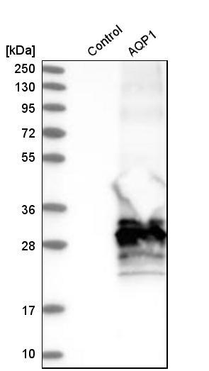
- Recombinant expression validation
- Main image
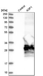
- Experimental details
- Western blot analysis in control (vector only transfected HEK293T lysate) and aQP1 over-expression lysate (Co-expressed with a C-terminal myc-DDK tag (~3.1 kDa) in mammalian HEK293T cells, LY403670).
- Sample type
- Human
- Protocol
- Protocol
Supportive validation
- Submitted by
- Atlas Antibodies (provider)
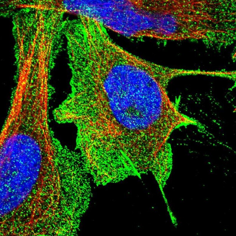
- Main image

- Experimental details
- Immunofluorescent staining of human cell line U-2 OS shows localization to plasma membrane.
- Sample type
- Human
Supportive validation
- Submitted by
- Atlas Antibodies (provider)
- Enhanced method
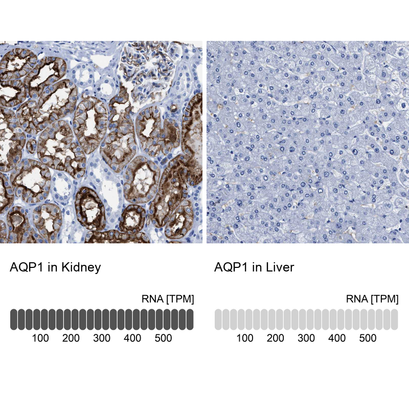
- Orthogonal validation
- Main image
- Experimental details
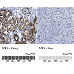
- Immunohistochemistry analysis in human kidney and liver tissues using HPA019206 antibody. Corresponding AQP1 RNA-seq data are presented for the same tissues.
- Sample type
- Human
- Protocol
- Protocol