Explore
 Validate
 Learn
Learn Western blot
 Immunocytochemistry
Antibody data
- Antibody Data
- Antigen structure
- References [3]
- Comments [0]
- Validations
- Immunocytochemistry [3]
- Immunohistochemistry [4]
Submit
Validation data
Reference
Comment
Report error
- Product number
- 14-9747-82 - Provider product page

- Provider
- Invitrogen Antibodies
- Product name
- Desmin Monoclonal Antibody (DE-U-10), eBioscience™
- Antibody type
- Monoclonal
- Antigen
- Other
- Description
- Description: The monoclonal antibody DE-U-10 recognizes desmin. Desmin is the protein subunit of class-III intermediate filaments. Desmin is found predominantly in skeletal, cardiac, and smooth muscle. Desmin plays a role in muscle cell development and differentiation, muscle cell architecture and structure, and mitochondrial function. Desmin forms scaffolds around the Z-disk of sarcomeres in muscle cells. Desmin knockout mice exhibit defects in skeletal, smooth, and cardiac muscle and have impaired mitochondrial function. Mutations in Desmin result in conditions such as desmin-related myopathy (DRM), cardiomyopathy dilated type 1I (CMD1I), and neurogenic scapuloperoneal syndrome Kaeser type (Kaeser syndrome). The DE-U-10 antibody recognizes human, mouse, rat, feline, bovine, goat, hamster, sheep, porcine, rabbit, and chicken desmin. The DE-U-10 antibody will also label tumors derived from muscle tissue, such as leiomyomas and rhabdomyosarcomas. Applications Reported: This DE-U-10 antibody has been reported for use in western blotting, immunohistochemical staining of formalin-fixed paraffin embedded tissue sections, microscopy, and immunocytochemistry. Applications Tested: This DE-U-10 antibody has been tested by immunohistochemistry of formalin-fixed paraffin embedded human tissue using either low or high pH antigen retrieval and can be used at less than or equal to 5 µg/mL. This DE-U-10 antibody has been tested by immunocytochemistry of methanol-fixed and permeabilized mouse cells and can be used at less than or equal to 5 µg/mL. It is recommended that the antibody be carefully titrated for optimal performance in the assay of interest. Purity: Greater than 90%, as determined by SDS-PAGE. Aggregation: Less than 10%, as determined by HPLC. Filtration: 0.2 µm post-manufacturing filtered.
- Reactivity
- Human, Mouse, Rat, Bovine, Chicken/Avian, Feline, Goat, Hamster, Porcine, Rabbit
- Host
- Mouse
- Isotype
- IgG
- Antibody clone number
- DE-U-10
- Vial size
- 100 μg
- Concentration
- 0.5 mg/mL
- Storage
- 4°C
Submitted references Tragedy in a heartbeat: malfunctioning desmin causes skeletal and cardiac muscle disease.
Desmin cytoskeleton linked to muscle mitochondrial distribution and respiratory function.
Desmin is essential for the tensile strength and integrity of myofibrils but not for myogenic commitment, differentiation, and fusion of skeletal muscle.
Goldfarb LG, Dalakas MC
The Journal of clinical investigation 2009 Jul;119(7):1806-13
The Journal of clinical investigation 2009 Jul;119(7):1806-13
Desmin cytoskeleton linked to muscle mitochondrial distribution and respiratory function.
Milner DJ, Mavroidis M, Weisleder N, Capetanaki Y
The Journal of cell biology 2000 Sep 18;150(6):1283-98
The Journal of cell biology 2000 Sep 18;150(6):1283-98
Desmin is essential for the tensile strength and integrity of myofibrils but not for myogenic commitment, differentiation, and fusion of skeletal muscle.
Li Z, Mericskay M, Agbulut O, Butler-Browne G, Carlsson L, Thornell LE, Babinet C, Paulin D
The Journal of cell biology 1997 Oct 6;139(1):129-44
The Journal of cell biology 1997 Oct 6;139(1):129-44
No comments: Submit comment
Supportive validation
- Submitted by
- Invitrogen Antibodies (provider)
- Main image
- Experimental details
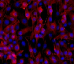
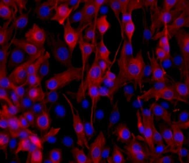
- Immunocytochemistry of methanol-fixed C2C12 cells using 5 µg/mL Anti-Desmin Purified, followed by F (ab')2 Anti-Mouse IgG eFluor® 570.Nuclei are stained with DAPI.
- Submitted by
- Invitrogen Antibodies (provider)
- Main image

- Experimental details
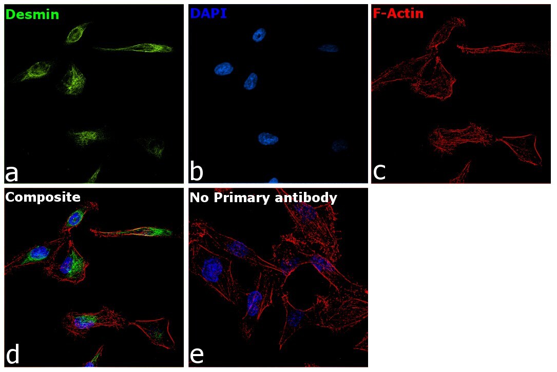
- Immunofluorescence analysis Desmin was performed using 70% confluent log phase RD cells. The cells were fixed with 4% paraformaldehyde for 10 minutes, permeabilized with 0.1% Triton™ X-100 for 15 minutes, and blocked with 2% BSA for 1 hour at room temperature. The cells were labeled with Desmin Monoclonal Antibody (DE-U-10), eBioscience™ (Product 14-9747-82) at 5 µg/mL in 0.1% BSA, incubated at 4 degree Celsius overnight and then with Goat anti-Mouse IgG (H+L), Superclonal™ Recombinant Secondary Antibody, Alexa Fluor 488 (Product # A28175) at a dilution of 1:2000 for 45 minutes at room temperature (Panel a: green). Nuclei (Panel b: blue) were stained with SlowFade® Gold Antifade Mountant with DAPI (Product # S36938). F-actin (Panel c: red) was stained with Rhodamine Phalloidin (Product # R415, 1:300). Panel d represents the merged image showing cytoskeletal localization. Panel e represents control cells with no primary antibody to assess background. The images were captured at 60X magnification.
- Submitted by
- Invitrogen Antibodies (provider)
- Main image

- Experimental details
- Immunofluorescence analysis Desmin was performed using 70% confluent log phase RD cells. The cells were fixed with 4% paraformaldehyde for 10 minutes, permeabilized with 0.1% Triton™ X-100 for 15 minutes, and blocked with 2% BSA for 1 hour at room temperature. The cells were labeled with Desmin Monoclonal Antibody (DE-U-10), eBioscience™ (Product 14-9747-82) at 5 µg/mL in 0.1% BSA, incubated at 4 degree Celsius overnight and then with Goat anti-Mouse IgG (H+L), Superclonal™ Recombinant Secondary Antibody, Alexa Fluor 488 (Product # A28175) at a dilution of 1:2000 for 45 minutes at room temperature (Panel a: green). Nuclei (Panel b: blue) were stained with SlowFade® Gold Antifade Mountant with DAPI (Product # S36938). F-actin (Panel c: red) was stained with Rhodamine Phalloidin (Product # R415, 1:300). Panel d represents the merged image showing cytoskeletal localization. Panel e represents control cells with no primary antibody to assess background. The images were captured at 60X magnification.

Supportive validation
- Submitted by
- Invitrogen Antibodies (provider)
- Main image
- Experimental details
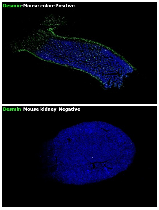
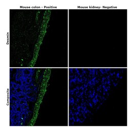
- Immunohistochemical analysis of Desmin was performed using formalin-fixed paraffin-embedded mouse colon and mouse kidney tissue sections. To expose the target protein, heat-induced epitope retrieval was performed on de-paraffinized sections using eBioscience™ IHC Antigen Retrieval Solution - High pH (10X) (Product # 00-4956-58) diluted to 1X solution in water in a decloaking chamber at 110 degree Celsius for 15 minutes. Following antigen retrieval, the sections were blocked with 2% normal goat serum in 1X PBS for 45 minutes at room temperature and then probed with Desmin Monoclonal Antibody (DE-U-10), eBioscience™ (Product # 14-9747-82) at 5 µg/mL in 0.1% normal goat serum overnight at 4 degree Celsius in a humidified chamber. Detection was performed using Goat anti-Mouse IgG (H+L) Highly Cross-Adsorbed Secondary Antibody, Alexa Fluor Plus 488 (Product # A32723) at a dilution of 1:2000 in 0.1% normal goat serum for 45 minutes at room temperature. Nuclei were stained with DAPI (Product # D1306) and the sections were mounted using ProLong™ Glass Antifade Mountant (Product # P36984). The images were captured on EVOS™ M7000 Imaging System (Product # AMF7000) at 20X magnification.

- Submitted by
- Invitrogen Antibodies (provider)
- Main image
- Experimental details
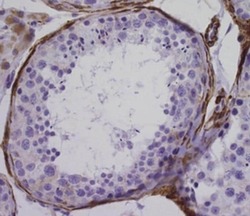
- Immunohistochemistry of formalin-fixed paraffin embedded human testes tissue using 5 µg/mL Anti-Desmin Purified, followed by Anti-Mouse IgG Biotin, Streptavidin HRP and DAB visualization.Nuclei are counterstained with hematoxylin.

- Submitted by
- Invitrogen Antibodies (provider)
- Main image
- Experimental details
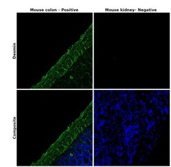
- Immunohistochemical analysis of Desmin was performed using formalin-fixed paraffin-embedded mouse colon and mouse kidney tissue sections. To expose the target protein, heat-induced epitope retrieval was performed on de-paraffinized sections using eBioscience™ IHC Antigen Retrieval Solution - High pH (10X) (Product # 00-4956-58) diluted to 1X solution in water in a decloaking chamber at 110 degree Celsius for 15 minutes. Following antigen retrieval, the sections were blocked with 2% normal goat serum in 1X PBS for 45 minutes at room temperature and then probed with Desmin Monoclonal Antibody (DE-U-10), eBioscience™ (Product # 14-9747-82) at 5 µg/mL in 0.1% normal goat serum overnight at 4 degree Celsius in a humidified chamber. Detection was performed using Goat anti-Mouse IgG (H+L) Highly Cross-Adsorbed Secondary Antibody, Alexa Fluor Plus 488 (Product # A32723) at a dilution of 1:2000 in 0.1% normal goat serum for 45 minutes at room temperature. Nuclei were stained with DAPI (Product # D1306) and the sections were mounted using ProLong™ Glass Antifade Mountant (Product # P36984). The images were captured on EVOS™ M7000 Imaging System (Product # AMF7000) at 20X magnification.

- Submitted by
- Invitrogen Antibodies (provider)
- Main image

- Experimental details
- Immunohistochemical analysis of Desmin was performed using formalin-fixed paraffin-embedded mouse colon and mouse kidney tissue sections. To expose the target protein, heat-induced epitope retrieval was performed on de-paraffinized sections using eBioscience™ IHC Antigen Retrieval Solution - High pH (10X) (Product # 00-4956-58) diluted to 1X solution in water in a decloaking chamber at 110 degree Celsius for 15 minutes. Following antigen retrieval, the sections were blocked with 2% normal goat serum in 1X PBS for 45 minutes at room temperature and then probed with Desmin Monoclonal Antibody (DE-U-10), eBioscience (Product # 14-9747-82) at 5 µg/mL in 0.1% normal goat serum overnight at 4 degree Celsius in a humidified chamber. Detection was performed using Goat anti-Mouse IgG (H+L) Highly Cross-Adsorbed Secondary Antibody, Alexa Fluor Plus 488 (Product # A32723) at a dilution of 1:2000 in 0.1% normal goat serum for 45 minutes at room temperature. Nuclei were stained with DAPI (Product # D1306) and the sections were mounted using ProLong™ Glass Antifade Mountant (Product # P36984). The images were captured on EVOS™ M7000 Imaging System (Product # AMF7000) at 20X magnification.