 Explore
 Validate
 Learn
Learn Western blot
Antibody data
- Antibody Data
- Antigen structure
- References [0]
- Comments [0]
- Validations
- Western blot [1]
- Immunohistochemistry [9]
Submit
Validation data
Reference
Comment
Report error
- Product number
- 50-9747-80 - Provider product page

- Provider
- Invitrogen Antibodies
- Product name
- Desmin Monoclonal Antibody (DE-U-10), eFluor™ 660, eBioscience™
- Antibody type
- Monoclonal
- Antigen
- Other
- Description
- Description: The monoclonal antibody DE-U-10 recognizes desmin. Desmin is the protein subunit of class-III intermediate filaments. Desmin is found predominantly in skeletal, cardiac, and smooth muscle. Desmin plays a role in muscle cell development and differentiation, muscle cell architecture and structure, and mitochondrial function. Desmin forms scaffolds around the Z-disk of sarcomeres in muscle cells. Desmin knockout mice exhibit defects in skeletal, smooth, and cardiac muscle and have impaired mitochondrial function. Mutations in Desmin result in conditions such as desmin-related myopathy (DRM), cardiomyopathy dilated type 1I (CMD1I), and neurogenic scapuloperoneal syndrome Kaeser type (Kaeser syndrome). The DE-U-10 antibody recognizes human, mouse, rat, feline, bovine, goat, hamster, sheep, porcine, rabbit, and chicken desmin. The DE-U-10 antibody will also label tumors derived from muscle tissue, such as leiomyomas and rhabdomyosarcomas. Applications Reported: This DE-U-10 antibody has been reported for use in immunohistochemical staining of formalin-fixed paraffin embedded tissue sections, microscopy, and immunocytochemistry. Applications Tested: This DE-U-10 antibody has been tested by immunohistochemistry of formalin-fixed paraffin embedded human tissue using high pH antigen retrieval and can be used at less than or equal to 10 µg/mL. This DE-U-10 antibody has been tested by immunocytochemistry of fixed and permeabilized cells and can be used at less than or equal to 10 µg/mL. It is recommended that the antibody be carefully titrated for optimal performance in the assay of interest. eFluor® 660 is a replacement for Alexa Fluor® 647. eFluor® 660 emits at 659 nm and is excited with the red laser (633 nm). Please make sure that your instrument is capable of detecting this fluorochome. Excitation: 633-647 nm; Emission: 668 nm; Laser: Red Laser. Filtration: 0.2 µm post-manufacturing filtered.
- Reactivity
- Human, Mouse, Rat, Bovine, Chicken/Avian, Feline, Goat, Hamster, Porcine, Rabbit
- Host
- Mouse
- Isotype
- IgG
- Antibody clone number
- DE-U-10
- Vial size
- 25 µg
- Concentration
- 0.2 mg/mL
- Storage
- 4° C, store in dark, DO NOT FREEZE!
No comments: Submit comment
Supportive validation
- Submitted by
- Invitrogen Antibodies (provider)
- Main image

- Experimental details
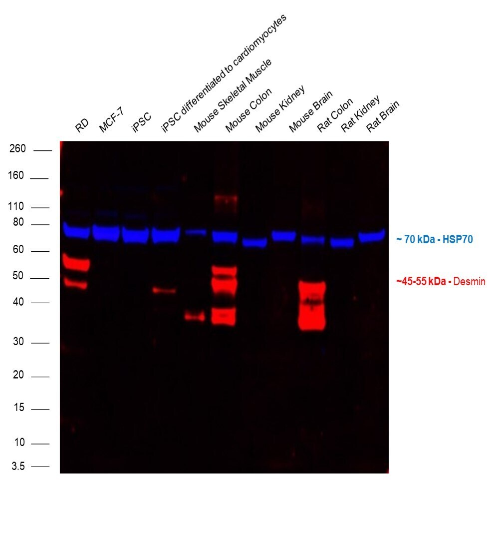
- Multiplexed fluorescent western blot was performed using Anti-Desmin Monoclonal Antibody (DE-U-10), eFluor™ 660, eBioscience™ (Product # 50-9747-82) and a 45 to 55 kDa band corresponding to desmin was observed in RD, iPSC differentiated to cardiomyocytes, mouse skeletal muscle, mouse colon and rat colon and not in other cell lines and tissues. Whole cell extracts (30 µg lysate) of RD (Lane 1), MCF7 (Lane 2), iPSC (Lane 3), iPSC differentiated to cardiomyocytes (Lane 4) and tissue extracts (30 µg lysate) of mouse skeletal muscle (Lane 5), mouse colon (Lane 6), mouse kidney (Lane 7), mouse brain (Lane 8), rat colon (Lane 9), rat kidney (Lane 10) and rat brain (Lane 11) were electrophoresed using NuPAGE™ 4-12% Bis-Tris Protein Gel (Product # NP0322BOX), 12 well. Resolved proteins were then transferred onto a nitrocellulose membrane (Product # IB23001) by iBlot® 2 Dry Blotting System (Product # IB21001). The blot was probed with the primary antibody Anti-Desmin Monoclonal Antibody (DE-U-10), eFluor™ 660, eBioscience™ (Product # 50-9747-82, 1 µg/mL) and HSP70 Polyclonal Antibody (Product # PA5-28003, 1:4000 dilution). Secondary antibody Donkey anti-Rabbit IgG (H+L) Highly Cross-Adsorbed Secondary Antibody, Alexa Fluor™ Plus 800 (Product # A32808, 1:10000 dilution) was used for detection of HSP70. Fluorescent detection was performed using iBright FL1500 (Product # A44115).
Supportive validation
- Submitted by
- Invitrogen Antibodies (provider)
- Main image

- Experimental details
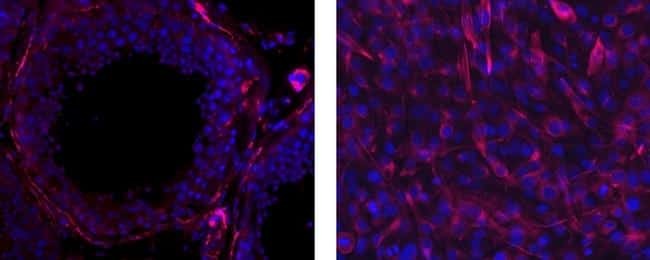
- Immunohistochemistry of formalin-fixed paraffin embedded human testes tissue using 10 µg/mL Anti-Desmin eFluor® 660, nuclei are stained with DAPI (left). Immunocytochemistry of methanol-fixed C2C12 cells using 10 µg/mL Anti-Desmin eFluor® 660, nuclei are stained with DAPI (right).
- Submitted by
- Invitrogen Antibodies (provider)
- Main image
- Experimental details
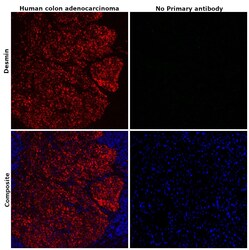
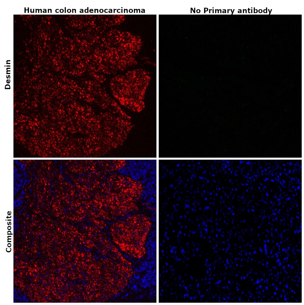
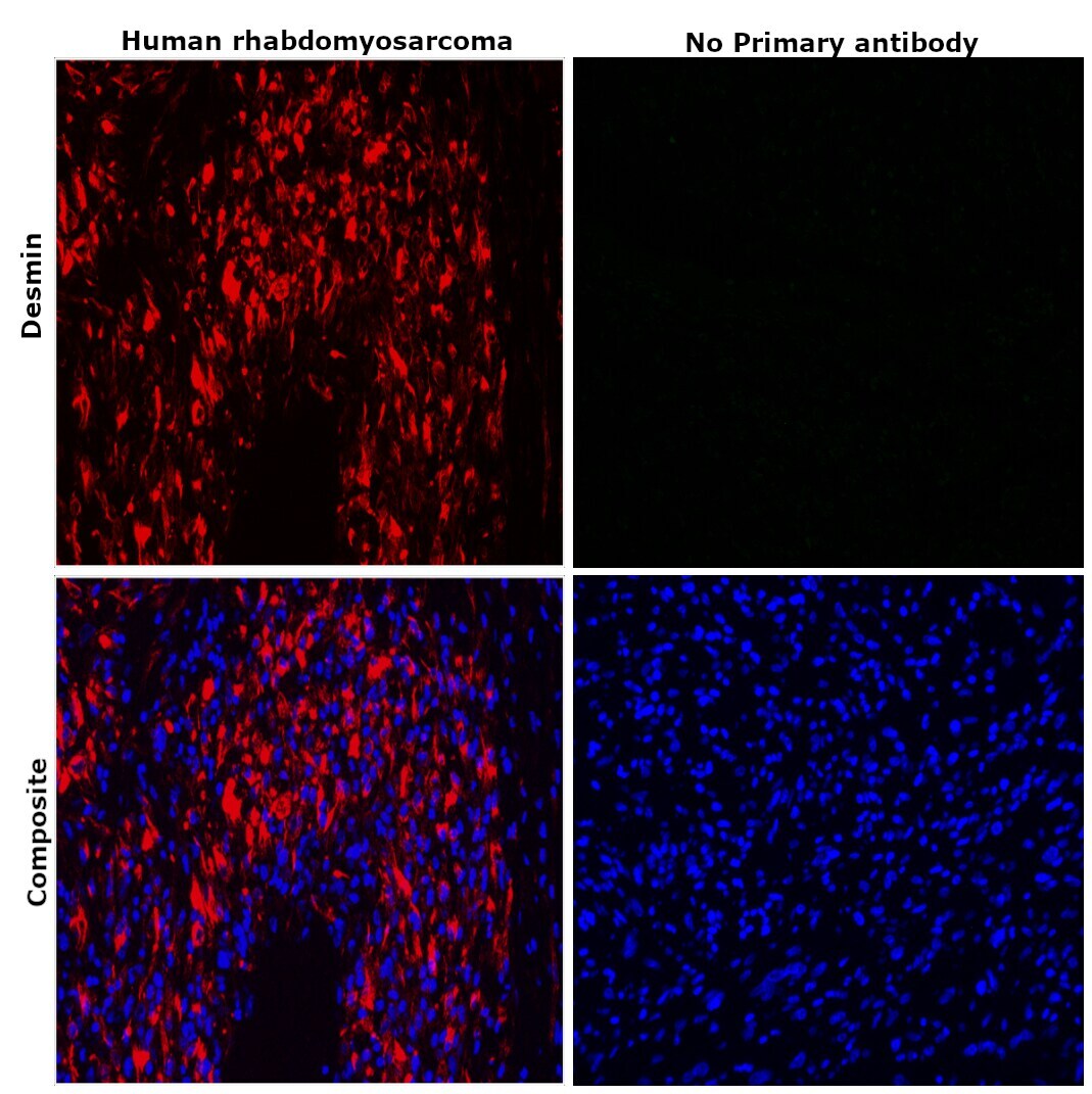
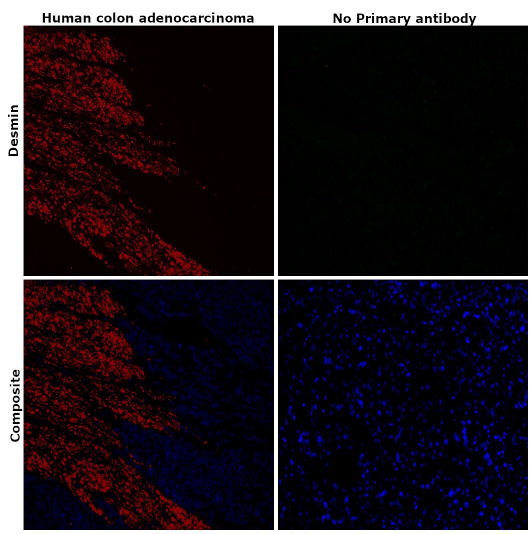
- Immunohistochemical analysis of desmin was performed using formalin-fixed paraffin-embedded human colon adenocarcinoma tissue sections. To expose the target protein, heat-induced epitope retrieval was performed on de-paraffinized sections using eBioscience™ IHC Antigen Retrieval Solution - Low pH (10X) (Product # 00-4955-58) diluted to 1X solution in water in a decloaking chamber at 110 degree Celsius for 15 minutes. Following antigen retrieval, the sections were blocked with 2% normal goat serum in 1X PBS for 45 minutes at room temperature and then probed with or without Desmin Monoclonal Antibody (DE-U-10), eFluor™ 660, eBioscience™ (Product # 50-9747-82) at a concentration of 2 µg/mL in 0.1% normal goat serum overnight at 4 degree Celsius in a humidified chamber. ReadyProbes™ Tissue Autofluorescence Quenching Kit (Product # R37630) was used to quench autofluorescence from the tissues. Nuclei were stained with DAPI (Product # D1306) and the sections were mounted using ProLong™ Glass Antifade Mountant (Product # P36984). The images were captured on EVOS™ M7000 Imaging System (Product # AMF7000) at 20X magnification and externally deconvoluted.
- Submitted by
- Invitrogen Antibodies (provider)
- Main image
- Experimental details
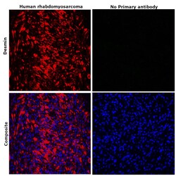
- Immunohistochemical analysis of desmin was performed using formalin-fixed paraffin-embedded human rhabdomyosarcoma tissue sections. To expose the target protein, heat-induced epitope retrieval was performed on de-paraffinized sections using eBioscience™ IHC Antigen Retrieval Solution - Low pH (10X) (Product # 00-4955-58) diluted to 1X solution in water in a decloaking chamber at 110 degree Celsius for 15 minutes. Following antigen retrieval, the sections were blocked with 2% normal goat serum in 1X PBS for 45 minutes at room temperature and then probed with or without Desmin Monoclonal Antibody (DE-U-10), eFluor™ 660, eBioscience™ (Product # 50-9747-82) at a concentration of 2 µg/mL in 0.1% normal goat serum overnight at 4 degree Celsius in a humidified chamber. ReadyProbes™ Tissue Autofluorescence Quenching Kit (Product # R37630) was used to quench autofluorescence from the tissues. Nuclei were stained with DAPI (Product # D1306) and the sections were mounted using ProLong™ Glass Antifade Mountant (Product # P36984). The images were captured on EVOS™ M7000 Imaging System (Product # AMF7000) at 20X magnification and externally deconvoluted.
- Submitted by
- Invitrogen Antibodies (provider)
- Main image

- Experimental details
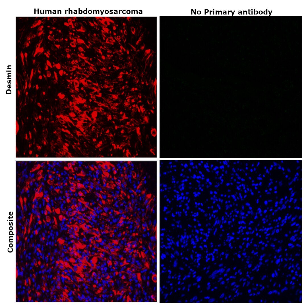
- Immunohistochemical analysis of desmin was performed using formalin-fixed paraffin-embedded human rhabdomyosarcoma tissue sections. To expose the target protein, heat-induced epitope retrieval was performed on de-paraffinized sections using eBioscience™ IHC Antigen Retrieval Solution - Low pH (10X) (Product # 00-4955-58) diluted to 1X solution in water in a decloaking chamber at 110 degree Celsius for 15 minutes. Following antigen retrieval, the sections were blocked with 2% normal goat serum in 1X PBS for 45 minutes at room temperature and then probed with or without Desmin Monoclonal Antibody (DE-U-10), eFluor™ 660, eBioscience™ (Product # 50-9747-82) at a concentration of 2 µg/mL in 0.1% normal goat serum overnight at 4 degree Celsius in a humidified chamber. ReadyProbes™ Tissue Autofluorescence Quenching Kit (Product # R37630) was used to quench autofluorescence from the tissues. Nuclei were stained with DAPI (Product # D1306) and the sections were mounted using ProLong™ Glass Antifade Mountant (Product # P36984). The images were captured on EVOS™ M7000 Imaging System (Product # AMF7000) at 20X magnification and externally deconvoluted.
- Submitted by
- Invitrogen Antibodies (provider)
- Main image
- Experimental details
- Immunohistochemical analysis of desmin was performed using formalin-fixed paraffin-embedded human colon adenocarcinoma tissue sections. To expose the target protein, heat-induced epitope retrieval was performed on de-paraffinized sections using eBioscience™ IHC Antigen Retrieval Solution - High pH (10X) (Product # 00-4956-58) diluted to 1X solution in water in a decloaking chamber at 110 degree Celsius for 15 minutes. Following antigen retrieval, the sections were blocked with 2% normal goat serum in 1X PBS for 45 minutes at room temperature and then probed with or without Desmin Monoclonal Antibody (DE-U-10), eFluor™ 660, eBioscience™ (Product # 50-9747-82) at a concentration of 2 µg/mL in 0.1% normal goat serum overnight at 4 degree Celsius in a humidified chamber. ReadyProbes™ Tissue Autofluorescence Quenching Kit (Product # R37630) was used to quench autofluorescence from the tissues. Nuclei were stained with DAPI (Product # D1306) and the sections were mounted using ProLong™ Glass Antifade Mountant (Product # P36984). The images were captured on EVOS™ M7000 Imaging System (Product # AMF7000) at 20X magnification and externally deconvoluted.
- Submitted by
- Invitrogen Antibodies (provider)
- Main image
- Experimental details
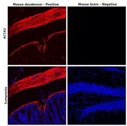
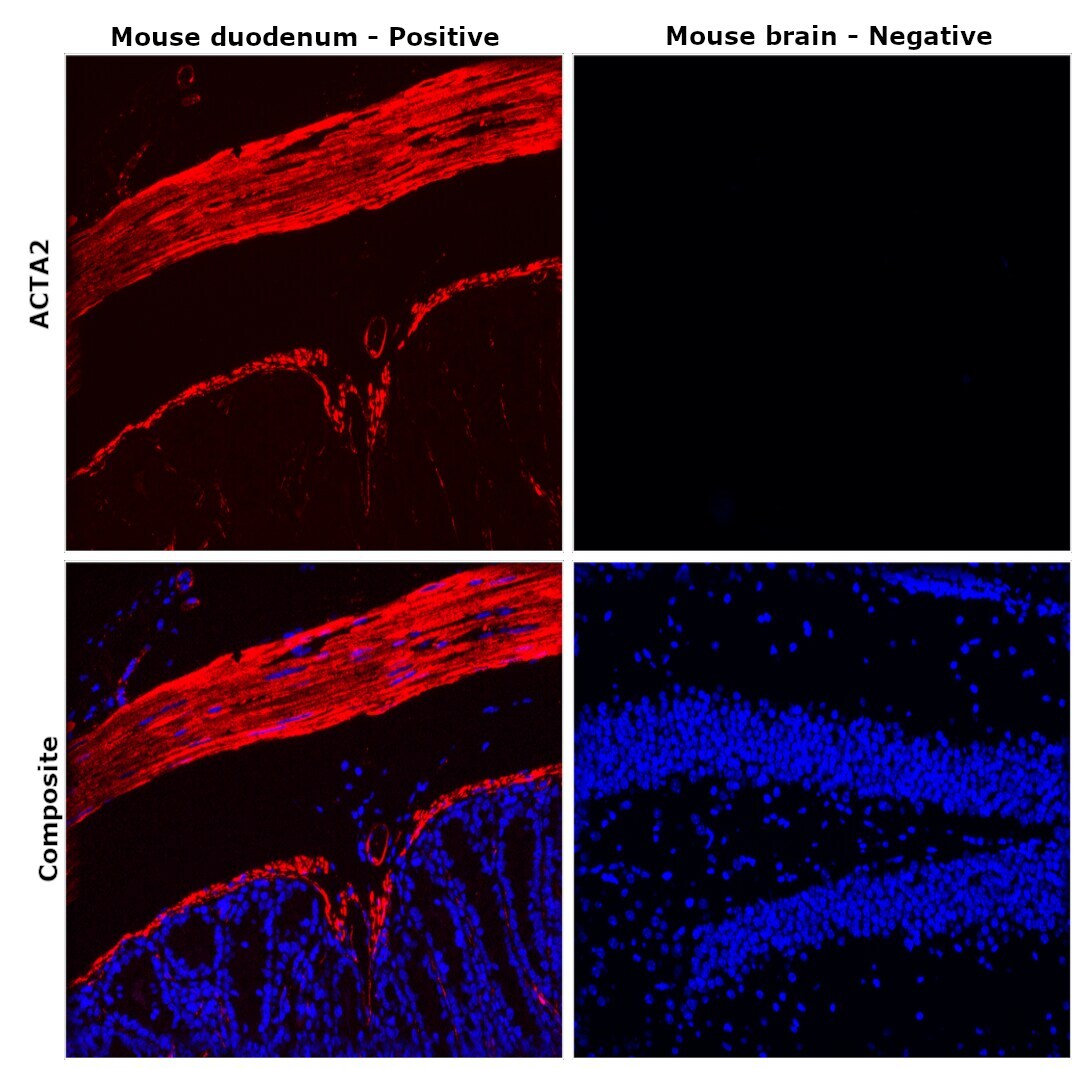
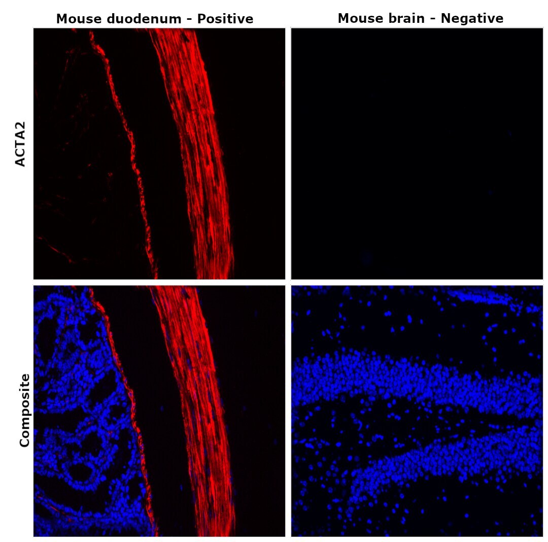
- Immunohistochemical analysis of Desmin was performed using formalin-fixed paraffin-embedded mouse duodenum and mouse brain tissue sections. To expose the target protein, heat-induced epitope retrieval was performed on de-paraffinized sections using eBioscience™ IHC Antigen Retrieval Solution - Low pH (10X) (Product # 00-4955-58) diluted to 1X solution in water in a microwave at 100 degree Celsius for 10 minutes. Following antigen retrieval, the sections were blocked with 2% normal goat serum in 1X PBS for 45 minutes at room temperature and then probed with Desmin Monoclonal Antibody (DE-U-10), eFluor™ 660, eBioscience™ (Product # 50-9747-82) at a concentration of 2 µg/mL in 0.1% normal goat serum overnight at 4 degree Celsius in a humidified chamber. ReadyProbes™ Tissue Autofluorescence Quenching Kit (Product # R37630) was used to quench autofluorescence from the tissues. Nuclei were stained with DAPI (Product # D1306) and the sections were mounted using ProLong™ Glass Antifade Mountant (Product # P36984). The images were captured on EVOS™ M7000 Imaging System (Product # AMF7000) at 20X magnification and externally deconvoluted.
- Submitted by
- Invitrogen Antibodies (provider)
- Main image
- Experimental details
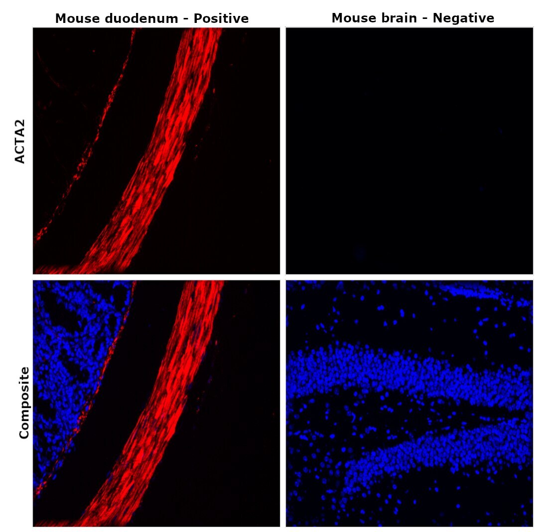
- Immunohistochemical analysis of Desmin was performed using formalin-fixed paraffin-embedded mouse duodenum and mouse brain tissue sections. To expose the target protein, heat-induced epitope retrieval was performed on de-paraffinized sections using eBioscience™ IHC Antigen Retrieval Solution - High pH (10X) (Product # 00-4956-58) diluted to 1X solution in water in a microwave at 100 degree Celsius for 10 minutes. Following antigen retrieval, the sections were blocked with 2% normal goat serum in 1X PBS for 45 minutes at room temperature and then probed with Desmin Monoclonal Antibody (DE-U-10), eFluor™ 660, eBioscience™ (Product # 50-9747-82) at a concentration of 2 µg/mL in 0.1% normal goat serum overnight at 4 degree Celsius in a humidified chamber. ReadyProbes™ Tissue Autofluorescence Quenching Kit (Product # R37630) was used to quench autofluorescence from the tissues. Nuclei were stained with DAPI (Product # D1306) and the sections were mounted using ProLong™ Glass Antifade Mountant (Product # P36984). The images were captured on EVOS™ M7000 Imaging System (Product # AMF7000) at 20X magnification and externally deconvoluted.
- Submitted by
- Invitrogen Antibodies (provider)
- Main image

- Experimental details
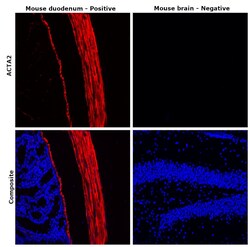
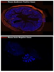
- Immunohistochemical analysis of Desmin was performed using formalin-fixed paraffin-embedded mouse duodenum and mouse brain tissue sections. To expose the target protein, heat-induced epitope retrieval was performed on de-paraffinized sections using eBioscience™ IHC Antigen Retrieval Solution - High pH (10X) (Product # 00-4956-58) diluted to 1X solution in water in a microwave at 100 degree Celsius for 10 minutes. Following antigen retrieval, the sections were blocked with 2% normal goat serum in 1X PBS for 45 minutes at room temperature and then probed with Desmin Monoclonal Antibody (DE-U-10), eFluor™ 660, eBioscience™ (Product # 50-9747-82) at a concentration of 2 µg/mL in 0.1% normal goat serum overnight at 4 degree Celsius in a humidified chamber. ReadyProbes™ Tissue Autofluorescence Quenching Kit (Product # R37630) was used to quench autofluorescence from the tissues. Nuclei were stained with DAPI (Product # D1306) and the sections were mounted using ProLong™ Glass Antifade Mountant (Product # P36984). The images were captured on EVOS™ M7000 Imaging System (Product # AMF7000) at 20X magnification and externally deconvoluted.
- Submitted by
- Invitrogen Antibodies (provider)
- Main image
- Experimental details
- Immunohistochemical analysis of Desmin was performed using formalin-fixed paraffin-embedded mouse duodenum and mouse brain tissue sections. To expose the target protein, heat-induced epitope retrieval was performed on de-paraffinized sections using eBioscience™ IHC Antigen Retrieval Solution - High pH (10X) (Product # 00-4956-58) diluted to 1X solution in water in a microwave at 100 degree Celsius for 10 minutes. Following antigen retrieval, the sections were blocked with 2% normal goat serum in 1X PBS for 45 minutes at room temperature and then probed with Desmin Monoclonal Antibody (DE-U-10), eFluor™ 660, eBioscience™ (Product # 50-9747-82) at a concentration of 2 µg/mL in 0.1% normal goat serum overnight at 4 degree Celsius in a humidified chamber. ReadyProbes™ Tissue Autofluorescence Quenching Kit (Product # R37630) was used to quench autofluorescence from the tissues. Nuclei were stained with DAPI (Product # D1306) and the sections were mounted using ProLong™ Glass Antifade Mountant (Product # P36984). The images were captured on EVOS™ M7000 Imaging System (Product # AMF7000) at 20X magnification.
