 Explore
 Validate
 Learn
Learn Western blot
Antibody data
- Antibody Data
- Antigen structure
- References [0]
- Comments [0]
- Validations
- Western blot [1]
- Immunocytochemistry [1]
Submit
Validation data
Reference
Comment
Report error
- Product number
- AF6019 - Provider product page

- Provider
- R&D Systems
- Product name
- Anti-Human/Mouse/Rat NM23-H1/H2 Antigen Affinity-purified Polyclonal Antibody
- Antibody type
- Polyclonal
- Antigen
- E. coli-derived recombinant human NM23‑H1, Met1-Glu152
- Description
- Antigen Affinity-purified
- Reactivity
- Human, Mouse, Rat
- Host
- Sheep
- Antigen sequence
P15531- Isotype
- IgG
- Vial size
- 100 µg
No comments: Submit comment
Supportive validation
- Submitted by
- R&D Systems (provider)
- Main image
- Experimental details
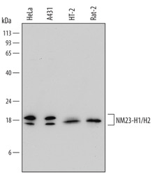
- Detection of Human, Mouse, and Rat NM23-H1/H2 by Western Blot. Western blot shows lysates of HeLa human cervical epithelial carcinoma cell line, A431 human epithelial carcinoma cell line, HT-2 mouse T cell line, and Rat-2 rat embryonic fibroblast cell line. PVDF Membrane was probed with 0.2 µg/mL of Human/Mouse/Rat NM23-H1/H2 Polyclonal Antibody (Catalog # AF6019) followed by HRP-conjugated Anti-Sheep IgG Secondary Antibody (Catalog # HAF016). Specific bands were detected for NM23-H1/H2 at approximately 17-20 kDa (as indicated). This experiment was conducted under reducing conditions and using Immunoblot Buffer Group 1.

Supportive validation
- Submitted by
- R&D Systems (provider)
- Main image
- Experimental details
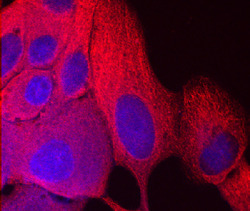
- NM23-H1/H2 in MCF-7 Human Cell Line. NM23-H1/H2 was detected in immersion fixed MCF-7 human breast cancer cell line using Sheep Anti-Human/Mouse/Rat NM23-H1/H2 Polyclonal Antibody (Catalog # AF6019) at 10 µg/mL for 3 hours at room temperature. Cells were stained using the NorthernLights™ 557-conjugated Anti-Sheep IgG Secondary Antibody (red; Catalog # NL010) and counterstained with DAPI (blue). Specific staining was localized to cytoplasm. View our protocol for Fluorescent ICC Staining of Cells on Coverslips.
