Explore
 Validate
 Learn
Learn Western blot
 Immunocytochemistry
Antibody data
- Antibody Data
- Antigen structure
- References [0]
- Comments [0]
- Validations
- Western blot [2]
- Immunohistochemistry [1]
- Flow cytometry [1]
Submit
Validation data
Reference
Comment
Report error
- Product number
- NBP2-34365-0.1mg - Provider product page

- Provider
- Novus Biologicals
- Product name
- Mouse Monoclonal GFAP Antibody
- Antibody type
- Monoclonal
- Description
- Protein G purified. This MAb recognizes a protein of ~50kDa which is identified as Glial Fibrillary Acidic Protein (GFAP). It shows no cross-reaction with other intermediate filament proteins. GFAP is specifically found in astroglia. GFAP is a very popular marker for localizing benign astrocyte and neoplastic cells of glial origin in the central nervous system. Antibody to GFAP is useful in differentiating primary gliomas from metastatic lesions in the brain and for documenting astrocytic differentiation in tumors outside the CNS.
- Reactivity
- Human, Mouse, Rat, Bovine, Chicken/Avian, Porcine, Rabbit
- Host
- Mouse
- Isotype
- IgG
- Vial size
- 0.1 mg
- Concentration
- 0.2 mg/ml
- Storage
- Store at 4C.
No comments: Submit comment
Supportive validation
- Submitted by
- Novus Biologicals (provider)
- Main image
- Experimental details
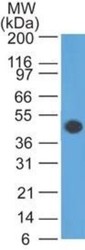
- Western Blot: GFAP Antibody (SPM507) [NBP2-34365] - Western blot analysis of GFAP in human brain lysate using GFAP Ab (SPM507).

- Submitted by
- Novus Biologicals (provider)
- Main image
- Experimental details
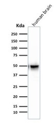
- Western Blot: GFAP Antibody (SPM507) [NBP2-34365] - Western Blot Analysis of human Brain tissue lysate using GFAP Antibody (SPM507).

Supportive validation
- Submitted by
- Novus Biologicals (provider)
- Main image
- Experimental details
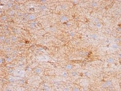
- Immunohistochemistry-Paraffin: GFAP Antibody (SPM507) [NBP2-34365] - Formalin-paraffin human brain stained with GFAP Ab (SPM507). Note cytoplasmic staining.

Supportive validation
- Submitted by
- Novus Biologicals (provider)
- Main image

- Experimental details
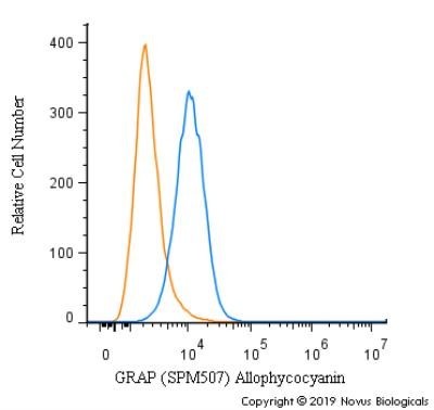
- Flow (Intracellular): GFAP Antibody (SPM507) [NBP2-34365] - An intracellular stain was performed on HeLa cells with U-87 [SPM507] Antibody NBP2-34413APC (blue) and a matched isotype control (orange). Cells were fixed with 4% PFA and then permeabilized with 0.1% saponin. Cells were incubated in an antibody dilution of 1 ug/mL for 30 minutes at room temperature. Both antibodies were conjugated to Allophycocyanin. Image using the Azide and BSA Free format of this antibody.