Explore
 Validate
 Learn
Learn Immunocytochemistry
 Immunohistochemistry
Antibody data
- Antibody Data
- Antigen structure
- References [3]
- Comments [0]
- Validations
- Immunocytochemistry [2]
- Flow cytometry [3]
- Other assay [1]
Submit
Validation data
Reference
Comment
Report error
- Product number
- 51-9792-82 - Provider product page

- Provider
- Invitrogen Antibodies
- Product name
- GFAP Monoclonal Antibody (2.2B10), Alexa Fluor™ 647, eBioscience™
- Antibody type
- Monoclonal
- Antigen
- Other
- Description
- Description: This 2.2B10 monoclonal antibody recognizes human, mouse, rat, and bovine glial fibrillary acid protein (GFAP). The immunogen used to generate this antibody was enriched bovine glial filaments. Applications Reported: This 2.2B10 antibody has been reported for use in intracellular staining followed by flow cytometric analysis. Applications Tested: This 2.2B10 antibody has been tested by intracellular staining followed by flow cytometric analysis of normal human peripheral blood cells using the Intracellular Fixation & Permeabilization Buffer Set (Product # 88-8824-00) and protocol. Please refer to Best Protocols: Protocol A: Two step protocol for (cytoplasmic) intracellular proteins located under the Resources Tab online. This may be used at less than or equal to 0.5 µg per test. A test is defined as the amount (µg) of antibody that will stain a cell sample in a final volume of 100 µL. Cell number should be determined empirically but can range from 10^5 to 10^8 cells/test. It is recommended that the antibody be carefully titrated for optimal performance in the assay of interest. Excitation: 633-647 nm; Emission: 668 nm; Laser: Red Laser.
- Reactivity
- Human, Mouse, Rat, Bovine
- Host
- Rat
- Conjugate
- Red dye
- Isotype
- IgG
- Antibody clone number
- 2.2B10
- Vial size
- 100 µg
- Concentration
- 0.2 mg/mL
- Storage
- 4° C, store in dark, DO NOT FREEZE!
Submitted references Immune-mediated neurodegenerative trait provoked by multimodal derepression of long-interspersed nuclear element-1.
In mouse chronic pancreatitis CD25(+)FOXP3(+) regulatory T cells control pancreatic fibrosis by suppression of the type 2 immune response.
Modulation of the IGF1R-MTOR pathway attenuates motor neuron toxicity of human ALS SOD1(G93A) astrocytes.
Takahashi F, Zhang C, Hohjoh H, Raveney B, Yamamura T, Hayashi N, Oki S
iScience 2022 May 20;25(5):104278
iScience 2022 May 20;25(5):104278
In mouse chronic pancreatitis CD25(+)FOXP3(+) regulatory T cells control pancreatic fibrosis by suppression of the type 2 immune response.
Glaubitz J, Wilden A, Golchert J, Homuth G, Völker U, Bröker BM, Thiele T, Lerch MM, Mayerle J, Aghdassi AA, Weiss FU, Sendler M
Nature communications 2022 Aug 3;13(1):4502
Nature communications 2022 Aug 3;13(1):4502
Modulation of the IGF1R-MTOR pathway attenuates motor neuron toxicity of human ALS SOD1(G93A) astrocytes.
Granatiero V, Sayles NM, Savino AM, Konrad C, Kharas MG, Kawamata H, Manfredi G
Autophagy 2021 Dec;17(12):4029-4042
Autophagy 2021 Dec;17(12):4029-4042
No comments: Submit comment
Supportive validation
- Submitted by
- Invitrogen Antibodies (provider)
- Main image

- Experimental details
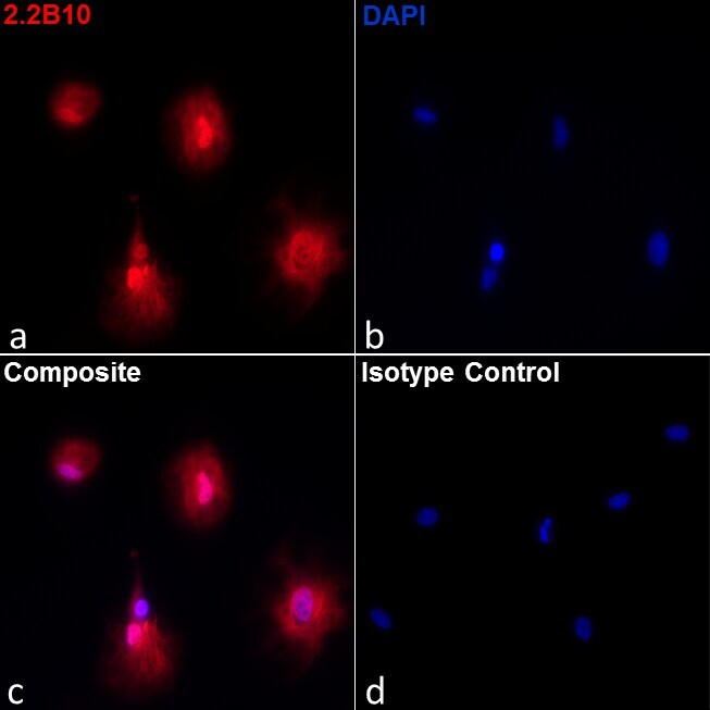
- Immunofluorescence analysis of GFAP Monoclonal Antibody, Alexa Fluor 647 (Product # 51-9792-82) on rat astrocytes. Rat primary cortical astrocytes were fixed with 4% paraformaldehyde for 15 minutes at room temperature, permeabilized with 1% Triton X-100 for 30 minutes at 37C, and blocked with ICC/IHC Blocking Buffer, Low Protein (Product # 00-4953-54) for 1 hour at room temperature. The cells were labeled with 10 µg/mL of GFAP Monoclonal Antibody, Alexa Fluor 647 (Product # 51-9792-82) in blocking buffer (Panel a: red). Nuclei (Panel b: blue) were stained with Fluoromount G with DAPI (Product # 00-4959). Panel c represents the merged image showing cytosolic and nuclear localization. Panel d represents a merged image of rat astrocytes labeled with 10 µg/mL of Rat IgG2a kappa Isotype Control, Alexa Fluor 647 (Product # 51-4321-81) antibody.
- Conjugate
- Red dye
- Submitted by
- Invitrogen Antibodies (provider)
- Main image
- Experimental details
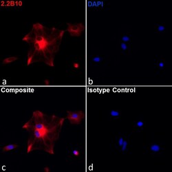
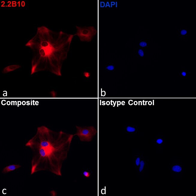
- Immunofluorescence analysis of GFAP Monoclonal Antibody, Alexa Fluor 647 (Product # 51-9792-82) on human primary astrocytes. The cells were fixed with 4% paraformaldehyde for 15 minutes at room temperature, permeabilized with 1% Triton X-100 for 30 minutes at 37C, and blocked with ICC/IHC Blocking Buffer, Low Protein (Product # 00-4953-54) for 1 hour at room temperature. Astrocytes were labeled with 10 µg/mL of GFAP Monoclonal Antibody, Alexa Fluor 647 (Product # 51-9792-82) in blocking buffer (Panel a: red). Nuclei (Panel b: blue) were stained with Fluoromount G with DAPI (Product # 00-4959). Panel c represents the merged image showing cytosolic localization. Panel d represents a merged image of human astrocytes labeled with 10 µg/mL of Rat IgG2a kappa Isotype Control, Alexa Fluor 647 (Product # 51-4321-81) antibody.
- Conjugate
- Red dye
Supportive validation
- Submitted by
- Invitrogen Antibodies (provider)
- Main image
- Experimental details
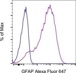
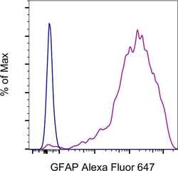
- U-251 cells were stained intracellularly, using the Intracellular Fixation & Permeabilization Buffer Set (Product # 88-8824-00) and protocol, with Rat IgG2a kappa Isotype Control, Alexa Fluor 647 (Product # 51-4321-81) (blue histogram) or GFAP Monoclonal Antibody, Alexa Fluor 647 (purple histogram). Total viable cells were used for analysis, as determined by Fixable Viability Dye eFluor 450 (Product # 65-0863-18).
- Conjugate
- Red dye
- Submitted by
- Invitrogen Antibodies (provider)
- Main image
- Experimental details
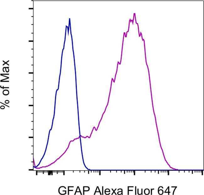
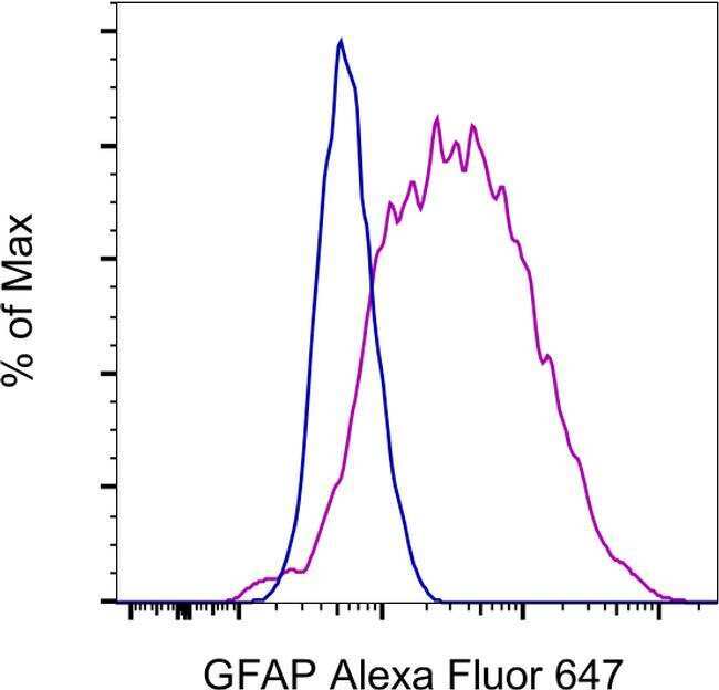
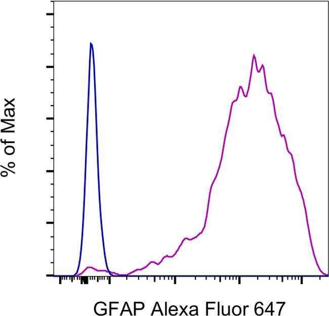
- Rat primary cortical astrocytes were stained intracellularly, using the Intracellular Fixation & Permeabilization Buffer Set (Product # 88-8824-00) and protocol, with Rat IgG2a kappa Isotype Control, Alexa Fluor 647 (Product # 51-4321-81) (blue histogram) or GFAP Monoclonal Antibody, Alexa Fluor 647 (purple histogram). Total viable cells were used for analysis, as determined by Fixable Viability Dye eFluor 450 (Product # 65-0863-18).
- Conjugate
- Red dye
- Submitted by
- Invitrogen Antibodies (provider)
- Main image
- Experimental details
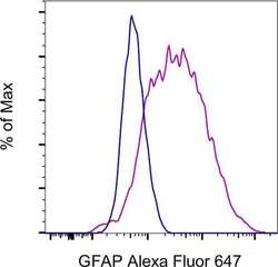
- Human astrocytes were stained intracellularly, using the Intracellular Fixation & Permeabilization Buffer Set (Product # 88-8824-00) and protocol, with Rat IgG2a kappa Isotype Control, Alexa Fluor 647 (Product # 51-4321-81) (blue histogram) or GFAP Monoclonal Antibody, Alexa Fluor 647 (purple histogram). Total viable cells were used for analysis, as determined by Fixable Viability Dye eFluor 450 (Product # 65-0863-18)
- Conjugate
- Red dye
Supportive validation
- Submitted by
- Invitrogen Antibodies (provider)
- Main image
- Experimental details
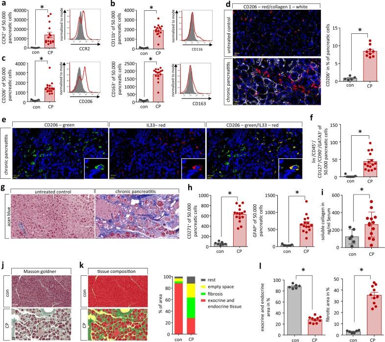
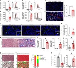
- Chronic pancreatitis leads to pancreatic tissue remodeling. a Flow cytometry analysis of isolated leukocytes from pancreatic tissue showed a significant increase of CCR2 + cells ( p = 0.0032, con n = 7/CP n = 15). b CD11b + labeling identified that also cells of the innate immune system infiltrate into the damaged pancreas ( p < 0.0001, con n = 7/CP n = 15). c Most of these cells were positive for CD206 ( p < 0.0001, con n = 7/CP n = 15) or CD163 ( p < 0.0001, con n = 7/CP n = 15), which characterized them as alternatively activated macrophages. d Labeling of CP tissue for CD206 illustrates their increase in pancreatic tissue of mice. A quantative analysis showed that ~9% of all cells were positive for CD206 in CP tissue ( p < 0.0001, con n = 5/CP n = 9), scale bars represent 20 um. e IL-33 expression of CD206 + macrophages was demonstrated by immunofluorescent labeling, scale bars represent 20 um. f This cytokine is a strong inducer of ILC2s (lin - CD45 + CD127 + CD90 + GATA3 + ) which were also significantly elevated in the pancreas of CP mice ( p = 0.0002, con n = 7/CP n = 15). g The replacement of exocrine pancreas by fibrotic tissue in CP was illustrated by azan blue staining of the pancreas, scale bars represent 50 um. h Flow cytometry analysis of isolated pancreatic cells for PSC marker proteins CD271 ( p < 0.0001, con n = 7/CP n = 15) and GFAP ( p < 0.0001, con n = 7/CP n = 15) identified a significant increase. i Serum collagen levels were significantly elevated in C
- Conjugate
- Red dye